
Grey matter
| Grey matter | |
|---|---|
 The formation of the spinal nerve from the dorsal and ventral roots (with grey matter labelled at centre right). | |
 Micrograph showing grey matter, with the characteristic neuronal cell bodies (dark shade of pink), and white matter with its characteristic fine meshwork-like appearance (left of image; lighter shade of pink). HPS stain. | |
| Details | |
| Identifiers | |
| Latin | substantia grisea |
| MeSH | D066128 |
| TA98 | A14.1.00.002 A14.1.02.020 A14.1.04.201 A14.1.05.201 A14.1.05.401 A14.1.06.301 |
| TA2 | 5365 |
| FMA | 67242 |
| Anatomical terminology | |
Grey matter, or brain matter in American English, is a major component of the central nervous system, consisting of neuronal cell bodies, neuropil (dendrites and unmyelinated axons), glial cells (astrocytes and oligodendrocytes), synapses, and capillaries. Grey matter is distinguished from white matter in that it contains numerous cell bodies and relatively few myelinated axons, while white matter contains relatively few cell bodies and is composed chiefly of long-range myelinated axons.[1] The colour difference arises mainly from the whiteness of myelin. In living tissue, grey matter actually has a very light grey colour with yellowish or pinkish hues, which come from capillary blood vessels and neuronal cell bodies.[2]
Structure
Grey matter refers to unmyelinated neurons and other cells of the central nervous system. It is present in the brain, brainstem and cerebellum, and present throughout the spinal cord.
Grey matter is distributed at the surface of the
Grey matter in the spinal cord is known as the
-
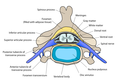 Cross-section of a spinal vertebra with the spinal cord in the centre (and grey matter labelled).
 -
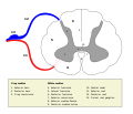 Cross-section of spinal cord with the grey matter labelled.


Grey matter undergoes development and growth throughout childhood and adolescence.[3] Recent studies using cross-sectional neuroimaging have shown that by around the age of 8 the volume of grey matter begins to decrease.[4] However, the density of grey matter appears to increase as a child develops into early adulthood.[4] Males tend to exhibit grey matter of increased volume but lower density than that of females.[5]
Function
Grey matter contains most of the brain's neuronal cell bodies.[6] The grey matter includes regions of the brain involved in muscle control, and sensory perception such as seeing and hearing, memory, emotions, speech, decision-making, and self-control.
The grey matter in the spinal cord is split into three grey columns:
- The pyramidal tract. These cells are responsible for the movement of muscles.
- The dorsal column-medial lemniscus tract and the spinothalamic tract.
- The lateral grey column is the third column of the spinal cord.
The grey matter of the spinal cord can be divided into different layers, called Rexed laminae. These describe, in general, the purpose of the cells within the grey matter of the spinal cord at a particular location.
-
 Interneurons present in the grey matter of the spinal cord
 -
 Rexed laminae groups the grey matter in the spinal cord according to its function.


Clinical significance
High
Meditation has been shown to change grey matter structure.[15][16][17][18][19]
Habitual playing of action video games has been reported to promote a reduction of grey matter in the hippocampus while 3D platformer games have been reported to increase grey matter in the hippocampus.[20][21][22]
Women and men with equivalent IQ scores have differing proportions of grey to white matter in cortical brain regions associated with intelligence.[23]
Pregnancy renders substantial changes in brain structure, primarily reductions in grey matter volume in regions subserving social cognition. The grey matter reductions endured for at least 2 years post-pregnancy.[24] The profile of brain changes is comparable to that taking place during adolescence, a hormonally similar transitional period of life.[25]
History
Etymology
In the current edition[26] of the official Latin nomenclature, Terminologia Anatomica, substantia grisea is used for English grey matter. The adjective grisea for grey is however not attested in classical Latin.[27] The adjective grisea is derived from the French word for grey, gris.[27] Alternative designations like substantia cana [28] and substantia cinerea[29] are being used alternatively. The adjective cana, attested in classical Latin,[30] can mean grey,[27] or greyish white.[31] The classical Latin cinerea means ash-coloured.[30]
Additional images
-
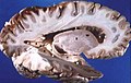 Human brain right dissected lateral view
 -
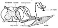
 Schematic representation of the chief ganglionic categories (I to V).


See also
References
- ISBN 978-0-87893-697-7.
- ISBN 978-0-7167-5300-1.
- PMID 11698594.
- ^ PMID 28432144.
- PMID 19906974.
- S2CID 23201991.
- S2CID 19928689.
- PMID 26072220.
- PMID 28646566.
- S2CID 22127231.
- PMID 15607838.
- PMID 18519827.
- S2CID 5968884.
- PMID 26602958.
- PMID 25632405.
- PMID 21071182.
- PMID 24643652.
- S2CID 207090878.
- PMID 19776221.
- PMID 25994669.
- "Playing action video games can actually harm your brain". Université de Montréal (Press release). 2017-08-07.
- ^ Collins K (10 August 2017). "Video games can either grow or shrink part of your brain, depending on how you play". qz.com. Archived from the original on 14 April 2018. Retrieved 5 May 2018.
- PMID 29211727.
- S2CID 4127512.
- S2CID 4113669.
- PMID 30663172.
- ^ Federative Committee on Anatomical Terminology (FCAT) (1998). Terminologia Anatomica. Stuttgart: Thieme[page needed]
- ^ a b c Triepel H (1910). Die anatomischen Namen. Ihre Ableitung und Aussprache. Mit einem Anhang: Biographische Notizen (3rd ed.). Wiesbaden: Verlag J.F. Bergmann.[page needed]
- ^ Triepel H (1910). Nomina Anatomica. Mit Unterstützung von Fachphilologen. Wiesbaden: Verlag J.F. Bergmann.[page needed]
- ^ Schreger CH (1805). "Synonymia anatomica. Synonymik der anatomischen Nomenclatur". In Fürth (ed.). Bureau für Literatur.[page needed]
- ^ a b Lewis CT, Short C (1879). A Latin dictionary founded on Andrews' edition of Freund's Latin dictionary. Oxford: Clarendon Press.[page needed]
- ^ Stearn WT (1983). Charles D (ed.). Botanical Latin. History, grammar, syntax, terminology and vocabulary (3rd ed.). London: Newton Abbot.[page needed]
External links
- May 2010, Stephanie Pappas (24 May 2010). "Why Is Gray Matter Gray?". Live Science.
{{cite web}}: CS1 maint: numeric names: authors list (link)
| National | |
|---|---|
| Other | |