Streptococcus agalactiae
| Streptococcus agalactiae | |
|---|---|

| |
| Scientific classification | |
| Domain: | Bacteria |
| Phylum: | Bacillota |
| Class: | Bacilli |
| Order: | Lactobacillales |
| Family: | Streptococcaceae |
| Genus: | Streptococcus |
| Species: | S. agalactiae
|
| Binomial name | |
| Streptococcus agalactiae Lehmann and Neumann, 1896
| |
Streptococcus agalactiae (also known as group B streptococcus or GBS) is a
S. agalactiae is the most common human pathogen of streptococci belonging to group B of the Rebecca
The plural term group B streptococci (referring to the serotypes) and the singular term group B streptococcus (referring to the single species) are both commonly used synonymously with S. agalactiae even though S. halichoeri and S. pseudoporcinus are also group B Streptococci. These species test positive as group B, but are not frequently carried by humans, and only rarely cause disease.[5]
In general, GBS is a harmless commensal bacterium being part of the human

S. agalactiae is also a common veterinary pathogen, because it can cause bovine mastitis (inflammation of the udder) in dairy cows. The species name agalactiae meaning "of no milk", alludes to this.[7]


Laboratory identification
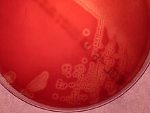
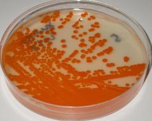
GBS grows readily on blood
GBS colonization
GBS is a normal component of the intestinal and vaginal microbiota in some women, GBS is an asymptomatic (presenting no symptoms) colonizer of the gastrointestinal tract and vagina in up to 30% of otherwise healthy adults, including pregnant women.[3][15] GBS colonization may be permanent, intermittent or temporary. In different studies, GBS vaginal colonization rate ranges from 0% to 36%, most studies reporting colonization rates in sexually active women over 20%.[16] It has been estimated that maternal GBS colonization worldwide is 18%, with regional variation from 11% to 35%.[17] These variations in the reported prevalence of asymptomatic GBS colonization could be related to the detection methods used, and differences in populations sampled.[15][18]
Virulence
As other virulent bacteria, GBS harbors an important number of virulence factors (virulence factors are molecules produced by bacteria that boosts their capacity to infect and damage human tissues), the most important being the capsular polysaccharide (rich in sialic acid)[3][19] and a pore-forming toxin, β-hemolysin.[19][20][21] Today it is considered that GBS pigment and hemolysin are identical or closely related molecules.[22][23][24][25]
Sialic acid is a notable virulence factor in S. agalactiae despite being found normally in humans and many other animals. By expressing an unusually high amount of sialic acid on the bacterial cell surface, S. agalactiae can subvert the
GBS infection in newborns
GBS colonization of the vagina usually does not cause problems in healthy women, nevertheless during pregnancy it can sometimes cause serious illness for the mother and the newborn. GBS is the leading cause of bacterial
GBS neonatal infection typically originates in the lower reproductive tract of infected mothers. GBS infections in newborns are separated into two clinical
Multistate surveillance 2006-2015 shows a decline in EOD from 0.37 to 0.23 per 1000 live births in the US but LOD remains steady at 0.31 per 1000 live births.[34]
It has been indicated that where there was a policy of providing IAP for GBS colonized mothers the overall risk of EOGBS is 0.3%.[35] Since 2006 to 2015 the incidence of GBS EOD decreased from 0.37 to 0.23 per thousand live births in the US.[36]
Though maternal GBS colonization is the key determinant for EOD, other factors also increase the risk. These factors include onset of labor before 37 weeks of gestation (
GBS LOD affects infants from 7 days to 3 months of age and is more likely to cause
Prevention of neonatal infection
The only reliable way to prevent EOD currently is intrapartum antibiotic prophylaxis (IAP), that is to say administration of antibiotics during delivery. It has been proved that intravenous penicillin or ampicillin administered for at least 4 hours before delivery to GBS colonized women is very effective at preventing vertical transmission of GBS from mother to baby and EOD. Intravenous penicillin remains the agent of choice for IAP, with intravenous ampicillin as an acceptable alternative.[3][30][31] For penicillin allergic women, the laboratory requisitions for ordering antepartum GBS screening cultures should indicate clearly the presence of penicillin allergy.[31] Cefazolin, clindamycin, and vancomycin are used to prevent EOD in infants born to penicillin-allergic mothers.[30][31] Intravenous vancomycin is recommended for IAP in women colonized with a clindamycin-resistant Group B Streptococcus strain and a severe penicillin allergy.[29][31]
There are two ways to identify female candidates to receive intrapartum antibiotic prophylaxis: a risk-based approach or a culture-based screening approach. The culture-based screening approach identifies candidates to receive IAP using lower vaginal and rectal cultures obtained between 36 and 37 weeks' gestation[30][31] (32–34 weeks of gestation for women with twins[41]) and IAP is administered to all GBS colonized women. The risk-based strategy identifies candidates to receive IAP by the aforementioned risk factors known to increase the probability of EOD without considering if the mother is or is not a GBS carrier.[3][42]
IAP is also recommended for women with intrapartum risk factors if their GBS carrier status is not known at the time of delivery, for women with GBS bacteriuria during their pregnancy, and for women who have had an infant with EOD previously.[citation needed]
The risk-based approach for IAP is in general less effective than the culture-based approach because in most of the cases EOD develops among newborns, which are born to mothers without risk factors.[18]
In 2010, the Centers for Disease Control and Prevention (CDC), in collaboration with several professional groups, issued its revised GBS prevention guidelines.[30]
In 2018, the task of revising and updating the GBS prophylaxis guidelines was transferred from the CDC [43] to ACOG (American College of Obstetricians and Gynecologists), the American Academy of Pediatrics and to the American Society for Microbiology.[14][29][31]
The ACOG committee issued an update document on Prevention of Group B Streptococcal Early-Onset Disease in Newborns in 2019.[31] This document does not introduce important changes from the CDC guidelines. The key measures necessary for preventing neonatal GBS early onset disease continue to be universal prenatal screening by culture of GBS from swabs collected from the lower vagina and rectum, correct collection and microbiological processing of the samples, and proper implementation of intrapartum antibiotic prophylaxis. The ACOG now recommends performing universal GBS screening between 36 and 37 weeks of gestation. This new recommendation provides a five-week window [44] for valid culture results that includes births that occur up to a gestational age of at least 41 weeks.
The culture-based screening approach is followed in most developed countries[45] such as the United States,[29][30][31] France,[46] Spain,[47] Belgium,[48] Canada, Argentina,[49] and Australia. The risk-based strategy is followed in the United Kingdom,[41][50] and the Netherlands.[18][51]
Screening for GBS colonization
Though the GBS colonization status of women can change during pregnancy, cultures to detect GBS carried out ≤5 weeks before delivery predict quite accurately the GBS carrier status at delivery.[citation needed]
In contrast, if the prenatal culture is performed more than five weeks before delivery it is unreliable for predicting accurately the GBS carrier status at delivery.[30][31][44][52][53]
The clinical specimens recommended for culture of GBS at 36–37 weeks' gestation, this recommendation provides a 5-week window for valid culture results that includes births that occur up to a gestational age of at least 41 weeks [31] (32–34 weeks of gestation for women with twins[41]) are swabs collected the lower vagina (near the introitus) and then from the rectum (through the anal sphincter) without use of a speculum.[30][31] Vaginal-rectal samples should be collected using a flocked swab preferably, since flocked swabs releases samples and microorganisms more effectively than fiber swabs.[14]
Following the recommendations of the
GBS-like colonies that develop in chromogenic media should be confirmed as GBS using additional reliable tests to avoid mis-identification.[9]
[30][31][41] Intrapartum NAAT without enrichment has a high false negative rate and the use of intrapartum NAAT without enrichment to rule out the need for IAP.[14]


Vaccination
Though IAP for EOD prevention is associated with a large decline in the incidence of the disease, there is, however, no effective strategy for preventing late-onset neonatal GBS disease.[54]
GBS infection in adults
GBS is also an important infectious agent able to cause invasive infections in adults. Serious life-threatening invasive GBS infections are increasingly recognized in the elderly and individuals compromised by underlying diseases such as diabetes, cirrhosis and cancer.[63] GBS infections in adults include urinary tract infection, skin and soft-tissue infection (skin and skin structure infection) bacteremia, osteomyelitis, meningitis and endocarditis.[6] GBS infection in adults can be serious and related with high mortality. In general penicillin is the antibiotic of choice for treatment of GBS infection.[64][65] Gentamicin (for synergy with penicillin G or ampicillin) can also be used in patients with life-threatening invasive GBS.[64]
Non-human infections
Streptococcus agalactiae was historically studied as a disease of cattle that harmed milk production, leading to its name "agalactiae" which means "absence of milk". Strains of bovine and human bacteria are generally interchangeable, with evidence of transmission from animals to humans and vice versa.[66]
Cattle
GBS is a major cause of mastitis (an infection of the udder) in dairy cattle and an important source of economic loss for the industry. GBS in cows can either produce an acute febrile disease or a subacute more chronic condition. Both lead to diminishing milk production (hence its name: agalactiae meaning "of no milk").[67] Outbreaks in herds are common, so this is of major importance for the dairy industry, and programs to reduce the impact of S. agalactiae disease have been enforced in many countries over the last 40 years.[7][66]
Other animals
GBS also causes severe epidemics in farmed fish, causing sepsis and external and internal hemorrhages, having been reported from wild and captive fish involved in
Vaccination is an effective method to prevent pathogenic diseases in aquaculture and different kinds vaccines to prevent GBS infections have been developed recently.[70]GBS has also been found in many other animals, such as camels, dogs, cats, crocodiles, seals, elephants and dolphins.[71][72]
References
- ^ ISBN 978-0-387-95041-9.
- ^ ISBN 978-0-8385-8529-0.
- ^ ISBN 978-0-443-06839-3.
- PMID 17634306.
- ^ "Guidelines for the Detection and Identification of Group B Streptococcus" (PDF). The American Society for Microbiology. July 23, 2021.
- ^ ISBN 978-0-443-06839-3.
- ^ PMID 9220132.
- ^ ISBN 978-0-323-08330-0.
- ^ PMID 28659318.
- PMID 16957264.)
{{cite journal}}: CS1 maint: multiple names: authors list (link - ^ PMID 10405420.
- PMID 30764872.
- PMID 20920213.
- ^ a b c d Filkins L, Hauser J, Robinson-Dunn Tibbetts R, Boyanton B, Revell P. "Guidelines for the Detection and Identification of Group B Streptococcus. March 10, 2020" (PDF). American Society for Microbiology. Archived from the original (PDF) on 27 June 2021. Retrieved 7 January 2021.
{{cite web}}: CS1 maint: multiple names: authors list (link) - ^ S2CID 25897076.
- PMID 25673591.
- PMID 29117327.
- ^ S2CID 15588906.
- ^ PMID 19257847.
- PMID 30711542.)
{{cite journal}}: CS1 maint: multiple names: authors list (link - PMID 32038561.)
{{cite journal}}: CS1 maint: multiple names: authors list (link - PMID 24617549.
- PMID 23712433.
- PMID 25750210.
- PMID 27383371.
- PMID 25400631.
- PMID 17768226.
- S2CID 11745321.
- ^ S2CID 195843897. Retrieved 7 January 2021.)
{{cite journal}}: CS1 maint: multiple names: authors list (link - ^ a b c d e f g h i j k l m n o Verani JR, McGee L, Schrag SJ (2010). "Prevention of perinatal group B streptococcal disease: revised guidelines from CDC, 2010" (PDF). MMWR Recomm Rep. 59(RR-10): 1–32.
- ^ S2CID 195659363.
- PMID 3931544.
- ^ CDC. "Group B Strep (GBS)-Clinical Overview". Retrieved 27 Oct 2015.
- PMID 30640366.)
{{cite journal}}: CS1 maint: multiple names: authors list (link - PMID 29117325.
- PMID 30640366.
- S2CID 15438484.
- S2CID 1013682.
- PMID 29117331.
- PMID 23973344.
- ^ PMID 28901693.
- S2CID 36906520.
- ^ CDC. "Prevention Guidelines. 2019 Guidelines Update". Retrieved 4 March 2021.
- ^ PMID 8885919.
- PMID 29117324.
- ^ Agence Nationale d’Accreditation et d’Evaluation en Santé. "Prévention anténatale du risque infectieux bactérien néonatal précoce. 2001" (PDF). Retrieved 22 December 2017.
- PMID 22488547. Retrieved 1 December 2019.
- ^ Belgian Health Council. "PREVENTION OF PERINATAL GROUP B STREPTOCOCCAL INFECTIONS. Guidelines from the Belgian Health Council, 2003" (PDF). Retrieved 22 December 2017.
- ^ Ministerio de Salud de la Nación. Dirección Nacional de Salud Materno Infantil. Argentina. "Recomendaciones para la prevención, diagnóstico y tratamiento de la infección neonatal precoz por Estreptococo β Hemolítico del Grupo B (EGB)" (PDF). Retrieved 2 December 2019.
- ^ RCOG and GBSS UK. "Group B Streptococcus (GBS) in pregnancy and newborn babies" (PDF). Retrieved 7 January 2021.
- PMID 17227807.)
{{cite journal}}: CS1 maint: multiple names: authors list (link - S2CID 26709882.
- S2CID 58106301.)
{{cite journal}}: CS1 maint: multiple names: authors list (link - S2CID 1533957.
- PMID 27767008.
- ^ PMID 26988258.
- PMID 31205250.
- ^ PMID 32425562.)
{{cite journal}}: CS1 maint: multiple names: authors list (link - PMID 24133184.
- PMID 23200934.
- PMID 27139805.
- PMID 29995578.
- PMID 30640366.)
{{cite journal}}: CS1 maint: multiple names: authors list (link - ^ PMID 16107984.
- PMID 11462195.
- ^ PMID 34486971.
- PMID 29153171. Retrieved 22 November 2019.
- PMID 19402966.
- PMID 24215651.
- PMID 28000606.
- PMID 23419028.
- PMID 28532793.
