
Portal vein
| Portal vein | |
|---|---|
pancreatic vein | |
| Drains to | liver sinusoid |
| Identifiers | |
| Latin | vena portae hepatis |
| MeSH | D011169 |
| TA98 | A12.3.12.001 |
| TA2 | 5092 |
| FMA | 50735 |
| Anatomical terminology] | |
The portal vein or hepatic portal vein (HPV) is a
The portal vein is not a true vein, because it conducts blood to capillary beds in the liver and not directly to the heart. It is a major component of the hepatic portal system, one of three portal venous systems in the human body; the others being the hypophyseal and renal portal systems.
The portal vein is usually formed by the confluence of the
Conditions involving the portal vein cause considerable illness and death. An important example of such a condition is elevated
Structure
Measuring approximately 8 cm (3 inches) long in adults,
In most individuals, the portal vein is formed by the union of the superior mesenteric vein and the splenic vein.[5] For this reason, the portal vein is occasionally called the splenic-mesenteric confluence.[4] Occasionally, the portal vein also directly communicates with the inferior mesenteric vein, although this is highly variable. Other tributaries of the portal vein include the cystic and the left and right gastric veins.[6] and also pararumbilical vein and prepyloric vein.
Immediately before reaching the liver, the portal vein divides into right and left. It ramifies further, forming smaller venous branches and ultimately portal venules. Each portal venule courses alongside a hepatic arteriole and the two vessels form the vascular components of the
Portacaval anastomoses
The
Accessory hepatic portal veins
Accessory hepatic portal veins are those veins that drain directly into the liver without joining the hepatic portal vein. These include the
Function
The portal vein and
Unlike most veins, the portal vein does not drain into the
Clinical significance
Portal hypertension
Increased blood pressure in the portal vein, called portal hypertension, is a major complication of liver disease, most commonly cirrhosis.[7] A dilated portal vein (diameter of greater than 13 or 15 mm) is a sign of portal hypertension, with a sensitivity estimated at 12.5% or 40%.[8] On Doppler ultrasonography, the main portal vein (MPV) peak systolic velocity normally ranges between 20 cm/s and 40 cm/s.[9] A slow velocity of <16 cm/s in addition to dilatation in the MPV are diagnostic of portal hypertension.[9]
Clinical
Pulsatility

Portal vein pulsatility can be measured by Doppler ultrasonography. An increased pulsatility may be caused by cirrhosis, as well as increased right atrial pressure (which in turn may be caused by right heart failure or tricuspid regurgitation).[9] Portal vein pulsatility can be quantified by pulsatility indices (PI), where an index above a certain cutoff indicates pathology:
| Index | Calculation | Cutoff |
|---|---|---|
| Average-based | (Max - Min) / Average[9] | 0.5[9] |
| Max-based | (Max - Min) / Max[11] | 0.5[11][12] - 0.54[12] |
Infection
Pylephlebitis is infection of the portal vein, usually arising from an infectious intra-abdominal process such as diverticulitis.[13][14]
Portal venous gas
Hepatic portal venous gas is a rare finding on radiological exams. Gas is shown to enter the portal venous system. It is most commonly caused by intestinal ischemia but has also been associated with colon cancer.[15]
Additional images
-
 Human embryo with heart and anterior body-wall removed to show the sinus venosus and its tributaries
 -
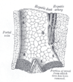
 Section across theportal triadof the pig
 -
 Longitudinal section of a small portal vein and canal
 -
 Hepatic portal vein. Plastination technique.
 -
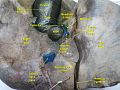
 Hepatic portal vein. Abdominal cavity. Deep dissection.
 -
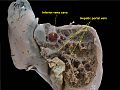
 Hepatic portal vein. Visceral surface of liver.


References
- S2CID 25169877.
- PMID 32818561.
- ISBN 978-0-7817-7174-0.
- ^ ISBN 978-3-540-65797-2.
- ISBN 978-1-55009-364-3.
- ^ a b c Henry Gray (1901). Anatomy, Descriptive and Surgical (16 ed.). Philadelphia: Lea Brothers. p. 619.
- ISBN 978-0-632-05582-1.
- PMID 19858596.
- ^ PMID 26169079.
- ISBN 978-1-85996-164-3.
- ^ ISSN 0195-668X.
- ^ ISBN 9783540938422.
- PMID 8589130.
- PMID 8488411.
- PMID 29390409.
External links
- Anatomy photo:38:12-0109 at the SUNY Downstate Medical Center - "Stomach, Spleen and Liver: The Visceral Surface of the Liver"
- Anatomy image:7959 at the SUNY Downstate Medical Center
- Anatomy image:8565 at the SUNY Downstate Medical Center
- Anatomy image:8697 at the SUNY Downstate Medical Center
- Cross section image: pembody/body8a—Plastination Laboratory at the Medical University of Vienna
- figures/chapter_30/30-2.HTM: Basic Human Anatomy at Dartmouth Medical School