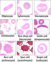
Hereditary stomatocytosis
| Hereditary stomatocytosis | |
|---|---|
 | |
| Stomatocytes | |
| Specialty | Hematology |

Hereditary stomatocytosis describes a number of inherited, mostly
Signs and symptoms
Stomatocytosis may present with signs and symptoms consistent with
Pathophysiology
The two varieties of stomatocytosis classified with respect to cellular hydration status are overhydrated (hydrocytosis) and dehydrated (xerocytosis).[2]: 225–226 Hereditary xerocytosis is characterized by autosomal dominant mutations in PIEZO1, which encodes a cation channel whose mechanosensitive properties enable erythrocytes to deform as they pass through narrow capillaries by decreasing their intracellular volume.[4] More rarely, hereditary xerocytosis may be caused by mutations in KCNN4, which encodes a calcium ion-sensitive potassium channel that mediates the potassium efflux triggered by a rise in intracellular Ca2+ via activated PIEZO1 channels.[4] Hereditary xerocytosis occurs more commonly in African populations,[2][4] and it exhibits complex interactions with other hereditary alterations of red blood cells, including sickle cell disease[4][5] and malaria resistance.[4][6]
Osmosis leads to the red blood cell having a constant tendency to swell and burst. This tendency is countered by manipulating the flow of sodium and potassium ions. A 'pump' forces sodium out of the cell and potassium in, and this action is balanced by a process called 'the passive leak'. In overhydrated hereditary stomatocytoses, the passive leak is increased and the erythrocyte becomes swamped with salt and water. The affected erythrocytes have increased osmotic fragility.
Overhydrated hereditary stomatocytosis is frequently linked to mutations in genes that encode components of the
Rare cases of hereditary spherocytosis can occur without cation leaks. These include cases of
Diagnosis
Ektacytometry may be helpful in distinguishing different subtypes of hereditary stomatocytosis.[4]
Variants
Haematologists have identified a number of variants. These can be classified as below.
- Overhydrated hereditary stomatocytosis[1]
- Dehydrated hereditary stomatocytosis (hereditary xerocytosis; hereditary hyperphosphatidylcholine haemolytic anaemia)[8]
- Dehydrated hereditary stomatocytosis with perinatal edema and/or pseudohyperkalemia[1]
- Cryohydrocytosis[1]
- 'Blackburn' variant[8]
- Familial pseudohyperkalaemia (not associated with hemolytic anemia)[1]
There are other families that do not fall neatly into any of these classifications.[9]
Stomatocytosis is also found as a hereditary disease in
Treatment
At present there is no specific treatment. Many patients with hemolytic anemia take
References
Further reading
- Eber SW, Lande WM, Iarocci TA, Mentzer WC, Höhn P, Wiley JS, Schröter W (July 1989). "Hereditary stomatocytosis: consistent association with an integral membrane protein deficiency". British Journal of Haematology. 72 (3): 452–455. S2CID 44935457.
- Hiebl-Dirschmied CM, Adolf GR, Prohaska R (June 1991). "Isolation and partial characterization of the human erythrocyte band 7 integral membrane protein". Biochimica et Biophysica Acta (BBA) - Biomembranes. 1065 (2): 195–202. PMID 1711899.
- Hiebl-Dirschmied CM, Entler B, Glotzmann C, Maurer-Fogy I, Stratowa C, Prohaska R (August 1991). "Cloning and nucleotide sequence of cDNA encoding human erythrocyte band 7 integral membrane protein". Biochimica et Biophysica Acta (BBA) - Gene Structure and Expression. 1090 (1): 123–124. PMID 1883838.
- Stewart GW, Hepworth-Jones BE, Keen JN, Dash BC, Argent AC, Casimir CM (March 1992). "Isolation of cDNA coding for an ubiquitous membrane protein deficient in high Na+, low K+ stomatocytic erythrocytes". Blood. 79 (6): 1593–1601. PMID 1547348.
