
Mandible
| Mandible | |
|---|---|
 Position of the mandible | |
 Animation of the mandible | |
| Details | |
| Precursor | First pharyngeal arch[1] |
| Identifiers | |
| Latin | mandibula |
| MeSH | D008334 |
| Anatomical terms of bone | |
In
The jawbone is the skull's only movable, posable bone, sharing joints with the cranium's temporal bones. The mandible hosts the lower teeth (their depth delineated by the alveolar process). Many muscles attach to the bone, which also hosts nerves (some connecting to the teeth) and blood vessels. Amongst other functions, the jawbone is essential for chewing food.
Owing to the
Structure
In humans, the mandible is the largest and lowest bone in the facial skeleton.[2] It is the only movable bone of the skull (discounting the vibrating ossicles of the middle ear).[3] It is connected to the skull's temporal bones by the temporomandibular joints. In addition to simply opening and closing, the jawbone can articulate side to side as well as forward and back.[4]
Components
The mandible consists of:
- The body, curving anteriorly like a horseshoe[5]
- Two rami (Latin: branch), rising from the posterior body, forming the (nearly square) angle of the mandible[5]
-
 Left side, lateral view (further from spine)
 -
 Left side, medial view (closer to spine)


Body


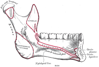
The body of the mandible is curved, and the front part gives structure to the
From the inside, the mandible appears concave. On either side of the lower symphysis is the

Borders
- The superior or alveolar border, wider behind than in front, is hollowed into cavities, for the reception of the teeth; these cavities are sixteen in number and vary in depth and size according to the teeth which they contain. To the outer lip of the superior border, on either side, the buccinator is attached as far forward as the first molartooth.
- The inferior border is rounded, longer than the superior, and thicker in front than behind; at the point where it joins the lower border of the ramus a shallow groove; for the facial artery, may be present.
Ramus
The ramus of the human mandible has four sides, two surfaces, four borders, and two processes. On the outside, the ramus is flat and marked by oblique ridges at its lower part. It gives attachment throughout nearly the whole of its extent to the masseter muscle.[8]
On the inside at the center there is an oblique
Borders
- The lower border of the ramus is thick, straight, and continuous with the inferior border of the body of the bone. At its junction with the posterior border is the angle of the mandible, which may be either inverted or everted and is marked by rough, oblique ridges on each side, for the attachment of the masseter laterally, and the medial pterygoid muscle medially; the stylomandibular ligament is attached to the angle between these muscles. The anterior border is thin above, thicker below, and continuous with the oblique line.[5]
- The region where the lower border meets the posterior border is the angle of the mandible.
- The posterior border is thick, smooth, rounded, and covered by the parotid gland. The upper border is thin, and is surmounted by two processes, the coronoid in front and the condyloid behind, separated by a deep concavity, the mandibular notch.[5]
Processes
- The coronoid process is a thin, triangular eminence, which is flattened from side to side and varies in shape and size.
- The condyloid process is thicker than the coronoid, and consists of two portions: the mandibular condyle, and the constricted portion which supports it, the neck. The condyle is the most superior part of the mandible and is part of the temporomandibular joint.[6]
- The mandibular notch, separating the two processes, is a deep semilunar depression and is crossed by the masseteric vessels and nerve.
Foramina
The mandible has two main holes (
- The mandibular foramen, is above the mandibular angle in the middle of each ramus.
- The mental foramen sits on either side of the mental protuberance (chin) on the body of mandible, usually inferior to the apices of the mandibular first and second premolars. As mandibular growth proceeds in young children, the mental foramen alters in direction of its opening from anterior to posterosuperior. The mental foramen allows the entrance of the mental nerve and blood vessels into the mandibular canal.[9]
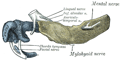
Nerves
The inferior alveolar nerve, a branch of the mandibular nerve (itself a major division of the cranium's trigeminal nerve), enters the mandibular foramen and runs forward in the mandibular canal, supplying sensation to the gums and teeth.[10] Before passing through the mental foramen, the nerve divides into two terminal branches: incisive and mental nerves. The incisive nerve runs forward in the mandible and supplies the anterior teeth. The mental nerve exits the mental foramen and supplies sensation to the chin and lower lip.[10]
Variation
Males generally have squarer, stronger, and larger mandibles than females. The mental protuberance is more pronounced in males but can be visualized and palpated in females.[citation needed]
Rarely, a bifid inferior alveolar nerve may be present, in which case a second mandibular foramen, more inferiorly placed, exists and can be detected by noting a doubled mandibular canal on a radiograph.[9]

Function
The mandible forms the lower jaw and holds the lower teeth in place. It articulates with the left and right temporal bones at the temporomandibular joints.
The condyloid process, the superior (upper) and posterior projection from the ramus, makes the temporomandibular joint with the temporal bone. The coronoid process, superior and anterior projection from the ramus. This provides attachment to the
Teeth sit in the upper part of the body of the mandible. The frontmost part of teeth is more narrow and holds front teeth. The back part holds wider and flatter (albeit grooved) teeth primarily for chewing food.[11] The word mandible derives from the Latin word mandibula 'jawbone' (literally, 'used for chewing'), from mandere 'to chew' and -bula (instrumental suffix).
In addition to mastication, the joint of the jawbone enables actions such speech and yawning,[12] while playing a more subtle role in activities such as kissing and breathing.[13]


Evolution
The mandible of vertebrates evolved from Meckel's cartilage, left and right segments of cartilage which supported the anterior branchial arch in early fish.[14]
In recent
Development
The mandible forms as a bone (
About the sixth week of fetal life, intramembranous ossification takes place in the membrane covering the outer surface of the ventral end of Meckel's cartilage, and each half of the bone is formed from a single center which appears, near the mental foramen.[5] By the tenth week, the portion of Meckel's cartilage which lies below and behind the incisor teeth is surrounded and invaded by the dermal bone (also known as the membrane bone). Somewhat later, accessory nuclei of cartilage make their appearance, as
- a wedge-shaped nucleus in the condyloid process and extending downward through the ramus;
- a small strip along the anterior border of the coronoid process;
- smaller nuclei in the front part of both alveolar walls and along the front of the lower border of the bone.[5]
These accessory nuclei possess no separate ossific centers but are invaded by the surrounding dermal bone and undergo absorption. The inner alveolar border, usually described as arising from a separate ossific center (splenial center), is formed in the human mandible by an ingrowth from the main mass of the bone.[5]
-
 Lateral (outer) view
 -
 Medial (inner) view


-
 Lateral (outer) view
 -
 Medial (inner) view


Aging
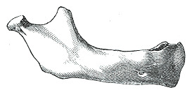
At birth, the body of the bone is a mere shell, containing the sockets of the two incisor, the canine, and the two deciduous
After birth, the two segments of the bone become joined at the symphysis, from below upward, in the first year; but a trace of separation may be visible in the beginning of the second year, near the alveolar margin. The body becomes elongated in its whole length, but more especially behind the mental foramen, to provide space for the three additional teeth developed in this part. The depth of the body increases owing to increased growth of the alveolar part, to afford room for the roots of the teeth, and by thickening of the subdental portion which enables the jaw to withstand the powerful action of the masticatory muscles; but, the alveolar portion is the deeper of the two, and, consequently, the chief part of the body lies above the oblique line. The mandibular canal, after the second dentition, is situated just above the level of the mylohyoid line; and the mental foramen occupies the position usual to it in the adult. The angle becomes less obtuse, owing to the separation of the jaws by the teeth; about the fourth year it is 140°.[5] The fibrocartilage of the mandibular symphysis fuses together in early childhood.[9]
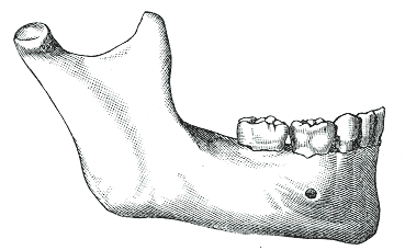
In the adult, the alveolar and subdental portions of the body are usually of equal depth. The mental foramen opens midway between the upper and lower borders of the bone, and the mandibular canal runs nearly parallel with the mylohyoid line. The ramus is almost vertical in direction, the angle measuring from 110° to 120°, also the adult condyle is higher than the coronoid process and the sigmoid notch becomes deeper.[5] The adult mandible is the skull's largest and strongest bone.[2]
In old age, the bone can become greatly reduced in volume where there is a loss of teeth, and consequent resorption of the alveolar process and interalveolar septa. Consequently, the chief part of the bone is below the oblique line. The mandibular canal, with the mental foramen opening from it, is closer to the alveolar border. The ramus is oblique in direction, the angle measures about 140°, and the neck of the condyle is more or less bent backward.[5]
-
 Newborn
 -
 Childhood
 -
 Adult
Adult -
 Old age


Clinical significance
Fracture

One fifth of
| Cause | Percentage |
|---|---|
| Motor vehicle accident | 40% |
| Assault | 10% |
| Fall | 10% |
| Sport | 5% |
| Other | 5% |
Dislocation and displacement
The mandible may be
The jawbone can also become deviated in mandibular lateral displacement, a condition which can offset facial symmetry and cause posterior crossbite.[21]
Resorption
The mandibular alveolar process can become resorbed when completely edentulous in the mandibular arch (occasionally noted also in partially edentulous cases). This resorption can occur to such an extent that the mental foramen is virtually on the superior border of the mandible, instead of opening on the anterior surface, changing its relative position. However, the more inferior body of the mandible is not affected and remains thick and rounded. With age and tooth loss, the alveolar process is absorbed so that the mandibular canal becomes nearer the superior border. Sometimes with excessive alveolar process absorption, the mandibular canal disappears entirely and leaves the inferior alveolar nerve without its bony protection, although it is still covered by soft tissue.[9]
Mandibulectomy
The surgical removal of all or part of the jawbone is known as a mandibulectomy.
Complications can involve difficulties with free flap transfer and airway management.[25][26] Additional side effects include pain, infection, numbness, and (rarely, fatal) bleeding.[27] Even successful surgeries can result in deformity, with an extreme version being referred to as the Andy Gump deformity after the comic book character, whose design apparently lacks a jaw; proposed reconstruction methods include implanting synthetic material, potentially involving 3D printing.[28]
Regeneration
Bone loss (as in osteoporosis) can be mitigated in the jawbone via bone grafting, which is sometimes performed to support dental implants (replacing teeth individually or in groups).[29]
Mandibular prosthetics date back to
In 2010, the first successful
Forensic medicine
The mandible can provide forensic evidence because its form changes over a person's life, and this can be used to determine a deceased person's age.[6]
Dental remains of
Other vertebrates
.jpg)
In
This complex primitive pattern has, however, been simplified to various degrees in the great majority of vertebrates, as bones have either fused or vanished entirely. In teleosts, only the dentary, articular, and angular bones remain, while in living amphibians, the dentary is accompanied only by the prearticular, and, in salamanders, one of the coronoids. The lower jaw of reptiles has only a single coronoid and splenial, but retains all the other primitive bones except the prearticular and the periosteum.[33]
While, in birds, these various bones have fused into a single structure, in mammals most of them have disappeared, leaving an enlarged dentary as the only remaining bone in the lower jaw – the mandible. As a result of this, the primitive jaw articulation, between the articular and quadrate bones, has been lost, and replaced with an entirely new articulation between the mandible and the temporal bone. An intermediate stage can be seen in some therapsids, in which both points of articulation are present. Aside from the dentary, only few other bones of the primitive lower jaw remain in mammals; the former articular and quadrate bones survive as the malleus and the incus of the middle ear.[33]
Finally, the
In culture
In the Hebrew Bible and Christian Old Testament Book of Judges, Samson used a donkey's jawbone to kill a thousand Philistines.[34]
As early as 1900, the phrase
Gobstoppers, a type of hard candy, are known in North America as jawbreakers due to the fracturing risk they impose on teeth.[36]
Owing in part to the forensic evidence of Hitler's death being limited to his dental remains (including a jawbone fragment broken and burnt around the alveolar process),[37] some fringe accounts (bolstered by the Soviet Union, which captured Berlin in 1945) allege that Hitler faked his death (ostensibly along with Eva Braun).[32]
In later decades, American real-estate businessman Fred Trump had a partial mandibulectomy which caused a conspicuous deformity.[38][39] In his fight against cancer, American film critic Roger Ebert had a partial mandibulectomy in 2006,[40] in addition to later surgeries.[31]
Additional images
-
 Cutaway view showingspongy bone
 -
 Turnaround with cranium
 -
 Anasal antrum and the maxillary sinuses.
_(14757789055).jpg)


See also
- Anatomical terms of location
- Bone terminology
- Mandible (arthropod mouthpart)
- Oral and maxillofacial surgery
- Simian shelf
References
![]() This article incorporates text in the public domain from page 172 of the 20th edition of Gray's Anatomy (1918)
This article incorporates text in the public domain from page 172 of the 20th edition of Gray's Anatomy (1918)
- ^ hednk-023—Embryo Images at University of North Carolina
- ^ a b Gray's Anatomy – The Anatomical Basis of Clinical Practice, 40th Edition, p. 530
- ISBN 9780470646083.
- Johns Hopkins Medicine. 8 August 2021. Retrieved 27 November 2023.
- ^ Lea & Febiger. pp. 172–177.
- ^ PMID 30335325, retrieved 8 July 2021
- ISBN 978-0-443-06684-9
- ISBN 978-0-683-06128-4.
- ^ a b c d Illustrated Anatomy of the Head and Neck, Fehrenbach and Herring, Elsevier, 2012, p. 59
- ^ PMID 31536318, retrieved 24 December 2023
- ^ "From Incisors to Molars:Tooth Types and Their Function". Main Street Smiles. Retrieved 4 January 2024.
- ^ a b "Temporomandibular Joint Disorder". Cedars-Sinai Medical Center. 2022. Retrieved 4 January 2024.
- ^ Deldar, Mike (22 January 2018). "6 Signs Your Partner Has TMJ: Tips from Indianapolis TMJ Specialist". Deldar Dental. Retrieved 4 January 2024.
- ^ a b "Palaeos Vertebrates: Bones: Gill Arches: Meckel's Cartilage". Palaeos. Retrieved 2 January 2024.
- ^ a b Pinhasi, R., Eshed, V., & von Cramon-Taubadel, N. (2015). Incongruity between affinity patterns based on mandibular and lower dental dimensions following the transition to agriculture in the Near East, Anatolia, and Europe. PLOS ONE, 10(2), e0117301.
- ^ Lieberman, D. E., Krovitz, G. E., Yates, F. W., Devlin, M., & Claire, M. S. (2004). Effects of food processing on masticatory strain and craniofacial growth in a retrognathic face. Journal of human evolution, 46(6), 655–677.
- ^ Pinhasi, R., Eshed, V., & Shaw, P. (2008). Evolutionary changes in the masticatory complex following the transition to farming in the southern Levant. American Journal of Physical Anthropology: The Official Publication of the American Association of Physical Anthropologists, 135(2), 136–148.
- ^ Kahn, S., Ehrlich, P., Feldman, M., Sapolsky, R., & Wong, S. (2020). The jaw epidemic: Recognition, origins, cures, and prevention. BioScience, 70(9), 759–771.
- ^ ]
- PMID 18634951.
- PMID 20362898.
- ^ "Mandibulectomy/Resection of the Jaw" (PDF). UAB Medicine. Retrieved 29 November 2023.
- ^ "Mandibulectomy: Definition, Procedure & Types". Cleveland Clinic. Retrieved 9 November 2023.
- ^ "Mandibulectomy". University of Illinois System. Retrieved 9 July 2023.
- S2CID 21807455.
- PMID 34976416.
- ^ "Mandibulectomy". SingHealth. Retrieved 12 January 2024.
- PMID 34401495.
- ^ "Jawbone Regeneration for Dental Implants". Harmony Dental Care. Retrieved 25 November 2023.
- PMID 1296501.
- ^ a b Ebert, Roger (6 May 2010). "Putting a better face on things". Roger Ebert. Retrieved 29 November 2023.
- ^ ISBN 978-1-85409-465-0.
- ^ ISBN 978-0-03-910284-5.
- ^ Judges 15:16 on BibleHub.
- ^ Liebenson, Donald (19 February 2020). "19 Classic Screwball Tex Avery Cartoons, Ranked From Best to Worst". Vulture. Retrieved 19 February 2024.
- ^ Project, CDHP Dental Health (22 August 2023). "Can Gobstoppers Break Your Teeth? (Everything You Need To Know)". CDHP Dental Health Project. Retrieved 11 March 2024.
- S2CID 29159362.and with our direct observations.
It is important to see that these data fit perfectly with the [Soviet] autopsy report
- ISBN 978-0393030297.
- ^ Travis, Abi (14 July 2020). "What Happened to Fred Trump's Face? The Internet Has a Few Theories". Distractify. Retrieved 28 November 2023.
- ^ Ebert, Roger (17 August 2006). "Email from Roger". RogerEbert.com. Archived from the original on 20 August 2006. Retrieved 18 January 2024.
External links
 Media related to Human mandible at Wikimedia Commons