
Hypoglossal nerve
| Hypoglossal nerve | |
|---|---|
thyrohyoid, intrinsic muscles of the tongue | |
| Identifiers | |
| Latin | nervus hypoglossus |
| MeSH | D007002 |
| NeuroNames | 704 |
| TA98 | A14.2.01.191 |
| TA2 | 6357 |
| FMA | 50871 |
| Anatomical terms of neuroanatomy] | |
| Cranial nerves |
|---|
|
The hypoglossal nerve, also known as the twelfth cranial nerve, cranial nerve XII, or simply CN XII, is a
The nerve is involved in controlling tongue movements required for speech and swallowing, including sticking out the tongue and moving it from side to side. Damage to the nerve or the neural pathways which control it can affect the ability of the tongue to move and its appearance, with the most common sources of damage being injury from trauma or surgery, and
Structure
The hypoglossal nerve arises as a number of small rootlets from the front of the
After emerging from the hypoglossal canal, the hypoglossal nerve gives off a meningeal branch and picks up a branch from the
At a point at the level of the
The rootlets of the hypoglossal nerve arise from the
-

-
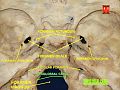 The hypoglossal nerve leaves the skull through the hypoglossal canal, which is situated near the large opening for the spinal cord, the foramen magnum.
 -
 After leaving the skull, the hypoglossal nerve spirals around the vagus nerve and then passes behind the deep belly of the digastric muscle.
 -
 The hypoglossal nerve then travelshyoglossus muscle, which it supplies. It then continues and supplies the genioglossusmuscle, and towards the tip of the tongue, where it divides into branches supplying the tongue muscles.


Development
Neurons of the hypoglossal nucleus are derived from the basal plate of the embryonic medulla oblongata.[7][8] The musculature they supply develops as the hypoglossal cord from the myotomes of the first four pairs of occipital somites.[9][10] The nerve is first visible as a series of roots in the fourth week of development, which have formed a single nerve and link to the tongue by the fifth week.[11][12]
Function

The hypoglossal nerve provides motor control of the extrinsic muscles of the tongue:
These muscles are involved in moving and manipulating the tongue.[2] The left and right genioglossus muscles in particular are responsible for protruding the tongue. The muscles, attached to the underside of the top and back parts of the tongue, cause the tongue to protrude and deviate towards the opposite side.[13] The hypoglossal nerve also supplies movements including clearing the mouth of saliva and other involuntary activities. The hypoglossal nucleus interacts with the reticular formation, involved in the control of several reflexive or automatic motions, and several corticonuclear originating fibers supply innervation aiding in unconscious movements relating to speech and articulation.[2]
Clinical significance
Damage
Reports of damage to the hypoglossal nerve are rare.
Examination
The hypoglossal nerve is tested by examining the tongue and its movements. At rest, if the nerve is injured a tongue may appear to have the appearance of a "bag of worms" (
The hypoglossal nerve carries lower motor neurons that synapse with upper motor neurons at the hypoglossal nucleus. Symptoms related to damage will depend on the position of damage in this pathway. If the damage is to the nerve itself (a lower motor neuron lesion), the tongue will curve toward the damaged side, owing to weakness of the genioglossus muscle of affected side which action is to deviate the tongue in the contralateral side .[19][20] If the damage is to the nerve pathway (an upper motor neuron lesion) the tongue will curve away from the side of damage, due to action of the affected genioglossus muscle, and will occur without fasciculations or wasting,[19] with speech difficulties more evident.[6] Damage to the hypoglossal nucleus will lead to wasting of muscles of the tongue and deviation towards the affected side when it is stuck out. This is because of the weaker genioglossal muscle.[2]
Use in nerve repair
The hypoglossal nerve may be connected (anastomosed) to the facial nerve to attempt to restore function when the facial nerve is damaged. Attempts at repair by either wholly or partially connecting nerve fibres from the hypoglossal nerve to the facial nerve may be used when there is focal facial nerve damage (for example, from trauma or cancer).[21][22]
Hypoglossal nerve stimulator implant
The hypoglossal nerve has also been clinically implicated in the treatment of obstructive sleep apnea.[23][24] Certain patients with obstructive sleep apnea who are deemed eligible candidates (e.g., failed continuous positive airway pressure therapy, underwent appropriate testing with drug induced sleep endoscopy, and meet other criteria as outlined by the FDA)[25] may be offered the hypoglossal nerve stimulator as an alternative. The purpose of the hypoglossal nerve stimulator is to relieve tongue base obstruction during sleep by stimulating the tongue to protrude during inspiration (i.e., inhale).
In this procedure, an electrical stimulator lead is placed around branches of the hypoglossal nerve that control tongue protrusion (e.g., genioglossus) via an incision in the neck.[26] A sensor lead is then placed in the chest between the ribs in the layer between the internal intercostal muscles and external intercostal muscles. The stimulator and sensory lead are then connected via a tunneled wire to an implantable pulse generator. When turned on during sleep, the sensory lead in the chest detects the respiratory cycle. During inspiration (i.e., inhale), an electrical signal is fired via the stimulator lead in the neck, stimulating the hypoglossal nerve, and causing the tongue to protrude, thereby alleviating obstruction.
History
The first recorded description of the hypoglossal nerve was by Herophilos (335–280 BC), although it was not named at the time. The first use of the name hypoglossal in Latin as nervi hypoglossi externa was used by Winslow in 1733. This was followed though by several different namings including nervi indeterminati, par lingual, par gustatorium, great sub-lingual by different authors, and gustatory nerve and lingual nerve (by Winslow). It was listed in 1778 as nerve hypoglossum magnum by Soemmering. It was then named as the great hypoglossal nerve by Cuvier in 1800 as a translation of Winslow and finally named in English by Knox in 1832.[27]
Other animals
The hypoglossal nerve is one of twelve cranial nerves found in amniotes including reptiles, mammals and birds.[28] As with humans, damage to the nerve or nerve pathway will result in difficulties moving the tongue or lapping water, decreased tongue strength, and generally cause deviation away from the affected side initially, and then to the affected side as contractures develop.[29] The evolutionary origins of the nerve have been explored through studies of the nerve in rodents and reptiles.[30] The nerve is regarded as arising evolutionarily from nerves of the cervical spine,[2] which has been incorporated into a separate nerve over the course of evolution.[30]
The size of the hypoglossal nerve, as measured by the size of the hypoglossal canal, has been hypothesised to be associated with the progress of evolution of primates, with reasoning that larger nerves would be associated with improvements in speech associated with evolutionary changes. This hypothesis has been refuted.[31]
See also
References
- ^ ISBN 978-0-87893-695-3.
- ^ ISBN 978-0-7020-4042-9.
- ISBN 978-0-323-04401-1.
- ^ a b c d Gray's Anatomy 2008, p. 460.
- ^ a b Gray's Anatomy 2008, p. 506-7.
- ^ ISBN 978-0-07-139011-8.
- ^ "Neural - Cranial Nerve Development". embryology.med.unsw.edu.au. Retrieved 17 June 2016.
- ^ Pansky, Ben. "Chapter 147. The Brainstem: Myelencephalon (fifth Vesicle) – Basal Motor Plate – Review of Medical Embryology Book – LifeMap Discovery". discovery.lifemapsc.com. Archived from the original on 13 March 2017. Retrieved 12 March 2017.
- ISBN 978-1455753604. Retrieved 12 March 2017.
- ISBN 9781607950325.
- ^ Hill, Mark. "Carnegie stage 12 – Embryology". embryology.med.unsw.edu.au. Retrieved 12 March 2017.
- PMID 6720613.
- ^ Gray's Anatomy 2008, p. 953.
- PMID 19494384.
- ^ PMID 8660159.
- ^ PMID 17622725.
- ^ a b "Chapter 7: Lower cranial nerves". www.dartmouth.edu. Archived from the original on 2007-10-18. Retrieved 2016-05-12.
- PMID 22296879.
- ^ a b c "Medical Neurosciences". Archived from the original on 2011-09-27. Retrieved 2011-12-04.
- ^ Brazis (2007). Localization in Clinical Neurology. p. 342.
- S2CID 36311886.
- ^ Ho, Tang. "Facial Nerve Repair Treatment". WebMDLLC. Retrieved 9 December 2011.
- PMID 33572156.
- S2CID 207937455.
- ^ "LCD - Hypoglossal Nerve Stimulation for the Treatment of Obstructive Sleep Apnea (L38307)". www.cms.gov. Retrieved 2024-01-01.
- ^ "Hypoglossal Nerve Stimulator Implantation (Selective Upper Airway Stimulation) | Iowa Head and Neck Protocols". medicine.uiowa.edu. Retrieved 2024-01-01.
- ISBN 978-0-19-534062-4.
- ^ Sharma, SK (2014). Objective Zoology. Krishna Prakashan Media. p. 3.84.
- ^ "Physical and Neurologic Examinations – Nervous System – Veterinary Manual". Veterinary Manual. Retrieved 2017-03-19.
- ^ PMID 26605049.
- ISBN 9780191009662.
- Sources
- Susan Standring; Neil R. Borley; et al., eds. (2008). Gray's anatomy : the anatomical basis of clinical practice (40th ed.). London: Churchill Livingstone. ISBN 978-0-8089-2371-8.
Notes
- ^ These are the genioglossus, hyoglossus, styloglossus, and intrinsic muscles of the tongue.