
Inferior mesenteric artery
| Inferior mesenteric artery | |
|---|---|
Large Intestine | |
| Identifiers | |
| Latin | arteria mesenterica inferior |
| MeSH | D017537 |
| TA98 | A12.2.12.069 |
| TA2 | 4291 |
| FMA | 14750 |
| Anatomical terminology] | |
In
Structure
Origin
The IMA arises from the anterior aspect of the abdominal aorta.[2][3]
Its origin is situated at the L3 vertebral level,[2][3] below the origins of the two renal arteries,[3] 3-4 cm above the aortic bifurcation,[3][2] at the level of the umbilicus, and posterior to the inferior border of the horizontal (III) part of the duodenum.[2]
Branches
Along its course, the IMA has the following branches:[1][4][3]
| Branch | notes |
| left colic artery | supplies descending colon |
| sigmoid branches | the most superior being described as 'the superior sigmoid artery' |
| superior rectal artery | effectively the terminal branch of the IMA (the continuation of the IMA after all other branches) |
All these arterial branches further divide into
Relations
The IMA is accompanied along its course by a similarly named vein, the inferior mesenteric vein, which drains into the splenic vein.[1] The IMV drains to the portal vein and does therefore not fully mirror the course of the IMA.[contradictory][1][4][3]
Distribution
Proximally, its territory of distribution overlaps (forms a
Clinical significance
The IMA and/or its branches must be resected for a left hemicolectomy.[5]
A horseshoe kidney, a common (1 in 500) anomaly of the kidneys, will be positioned below the IMA.[6][7]
Additional images
-
 The abdominal aorta and its branches.
 -
 The inferior mesenteric artery and its branches.
 -
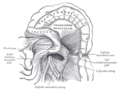
 Abdominal portion of the sympathetic trunk, with the celiac plexus and hypogastric plexus.
 -
 Duodenojejunal fossa.
 -
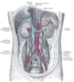 Posterior abdominal wall, after removal of the peritoneum, showing kidneys, suprarenal capsules, and great vessels.
 -
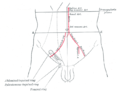 Front of abdomen, showing surface markings for arteries and inguinal canal.
 -
 Inferior mesenteric artery
 -
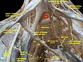
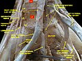
 Lumbar and sacral plexus. Deep dissection.Anterior view.
 -
 Lumbar and sacral plexus. Deep dissection.Anterior view.
 -
 Lumbar and sacral plexus. Deep dissection.Anterior view.


References
- ^ OCLC 920806541.
- ^ ISBN 978-0-7295-3752-0.
- ^ OCLC 881508489.
- ^ OCLC 813301028.
- PMID 27011519.
- PMID 25375751.
- ^ "Clinical case: Horseshoe kidney transplantation". Kenhub. Retrieved 2019-09-28.
External links
- Lotti M. Anatomy in relation to left colectomy
- Anatomy figure: 39:02-05 at Human Anatomy Online, SUNY Downstate Medical Center - "Branches of the inferior mesenteric artery."
- Anatomy photo:40:11-0103 at the SUNY Downstate Medical Center - "Posterior Abdominal Wall: Branches of the Abdominal Aorta"
- Anatomy image:7924 at the SUNY Downstate Medical Center
- Anatomy image:7997 at the SUNY Downstate Medical Center
- Anatomy image:8407 at the SUNY Downstate Medical Center
- Anatomy image:8659 at the SUNY Downstate Medical Center
- Atlas image: abdo_wall70 at the University of Michigan Health System - "Posterior Abdominal Wall, Dissection, Anterior View"
- sup&infmesentericart at The Anatomy Lesson by Wesley Norman (Georgetown University)