
Inferior vena cava
| Inferior vena cava | |
|---|---|
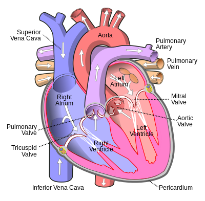 Anterior (frontal) view of the opened heart. White arrows indicate valid blood flow. | |
Right atrium | |
| Artery | abdominal aorta |
| Identifiers | |
| Latin | vena cava inferior |
| Acronym(s) | IVC |
| MeSH | D014682 |
| TA98 | A12.3.09.001 |
| TA2 | 4991 |
| FMA | 10951 |
| Anatomical terminology] | |
The inferior vena cava is a large vein that carries the deoxygenated
The inferior vena cava is the lower ("inferior") of the two venae cavae, the two large veins that carry deoxygenated blood from the body to the right atrium of the heart: the inferior vena cava carries blood from the lower half of the body whilst the superior vena cava carries blood from the upper half of the body. Together, the venae cavae (in addition to the coronary sinus, which carries blood from the muscle of the heart itself) form the venous counterparts of the aorta.
It is a large
Structure
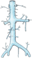
The IVC is formed by the joining of the left and right
The inferior vena cava begins as the left and right common iliac veins behind the
Tributaries
The specific levels of the tributaries are as follows:
| Level | Vein |
| T8 | hepatic veins, inferior phrenic vein |
| L1 | right renal veins
|
| L2 | right gonadal vein |
| L1–L5 | lumbar veins |
| L5 | common iliac veins
|
Because the inferior vena cava is located to the right of the midline, drainage of the tributaries is not always symmetrical. On the right, the
Development
In the
Variation
Rarely, the inferior vena cava may vary in its size and position. In
In between 0.2% to 0.3% of people,[5] the inferior vena cava may be duplicated beneath the level of the renal veins.[6]
Function
The inferior vena cava is a
The corresponding vein that carries deoxygenated blood from the upper half of the body is the superior vena cava.
Clinical significance
Health problems attributed to the IVC are most often associated with it being compressed (ruptures are rare because it has a low
Blockage of the inferior vena cava is rare and is treated urgently as a life-threatening condition. It is associated with

Trauma to the vena cava is usually fatal as unstoppable excessive blood loss occurs.
Additional images
-
 Inferior vena cava
 -
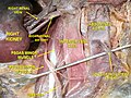 Inferior vena cava front view
 -
 Image of an inferior vena cava filter
 -
 Image showing an inferior vena cava filter in its position


See also
References
- ^ ISBN 978-0-12-369515-4, retrieved November 22, 2020
- ^ ISBN 978-1-4160-6839-6, retrieved November 22, 2020
- ^ ISBN 978-0-323-34062-5, retrieved November 22, 2020
- PMID 12484622.
- ISBN 978-0-323-06794-2, retrieved November 22, 2020
- ^ ISBN 978-0-8089-2371-8.
- S2CID 42135883.
- ^ Geehan DM, Inferior Vena Caval Thrombosis, emedicine.com, URL: http://www.emedicine.com/med/topic2718.htm, Accessed: August 3, 2005.
External links
- Anatomy photo:40:13-0101 at the SUNY Downstate Medical Center - "Posterior Abdominal Wall: Tributaries to the Inferior Vena Cava"
- Cross section image: pembody/body12a—Plastination Laboratory at the Medical University of Vienna