
Insect morphology
Insect morphology is the study and description of the
There is enormous variation in body structure amongst insect species. Individuals can range from 0.3 mm (fairyflies) to 30 cm across (great owlet moth);[1]: 7 have no eyes or many; well-developed wings or none; and legs modified for running, jumping, swimming, or even digging. These modifications allow insects to occupy almost every ecological niche except the deep ocean. This article describes the basic insect body and some variations of the different body parts; in the process, it defines many of the technical terms used to describe insect bodies.
Anatomy summary
Insects, like all arthropods, have no interior skeleton; instead, they have an
External
Exoskeleton
The insect's outer skeleton, the
From the embryonic stages, a layer of columnar or cuboidal epithelial cells gives rise to the external cuticle and an internal basement membrane. The majority of insect material is inside of the endocuticle. The cuticle provides muscular support and acts as a protective shield as the insect develops. However, since it cannot grow, the external sclerotized part of the cuticle is periodically shed in a process called "molting". As the time for molting approaches, most of the exocuticle material is reabsorbed. In molting, the old cuticle separates from the epidermis (apolysis). Enzymatic molting fluid is then released between the old cuticle and epidermis, which separates the exocuticle by digesting the endocuticle and sequestering its material for the new cuticle. When the new cuticle has formed sufficiently, the epicuticle and reduced exocuticle are shed in ecdysis.[5]: 16–20
The four principal regions of an insect body segment are the
Head


The head in most insects is enclosed in a hard, heavily sclerotized, exoskeletal head capsule. The main exception is in those species whose larvae are not fully sclerotized, mainly some holometabola; but even most unsclerotized or weakly sclerotized larvae tend to have well-sclerotized head capsules, for example, the larvae of Coleoptera and Hymenoptera. The larvae of Cyclorrhapha however, tend to have hardly any head capsule at all.
The head capsule bears most of the sensory organs, including the antennae, ocelli, and compound eyes, along with the mouthparts. In the adult insect, the head capsule appears unsegmented, though embryological studies show it to consist of six segments that bear the paired head appendages, including the mouthparts, each pair on a specific segment.[7] Each such pair occupies one segment, though not all segments in modern insects bear any visible appendages.
Of all the insect orders,
The ecdysial suture is made of the coronal, frontal, and epicranial sutures plus the ecdysial and cleavage lines, which vary among different species of insects. The ecdysial suture is longitudinally placed on the vertex, separating the epicranial halves of the head to the left and right sides. Depending on the insect, the suture may come in different shapes: like either a Y, U or V. Those diverging lines that make up the ecdysial suture are called the frontal or frontogenal sutures. Not all species of insects have frontal sutures, but in those that do, the sutures split open during ecdysis, which provides an opening for the new instar to emerge from the integument.
The

The
On the posterior aspect of the head are the
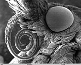
Compound eyes and ocelli
Most insects have one pair of large, prominent compound eyes composed of units called ommatidia (ommatidium, singular), up to 30,000 in a single compound eye of, for example, large dragonflies. This type of eye gives less resolution than eyes found in vertebrates, but it gives an acute perception of movement and usually possesses UV- and green sensitivity, and may have additional sensitivity peaks in other regions of the visual spectrum. Often an ability to detect the E-vector of polarized light exists in polarization of light.[11] There can also be an additional two or three ocelli, which help detect low light or small changes in light intensity. The image perceived is a combination of inputs from the numerous ommatidia, located on a convex surface, thus pointing in slightly different directions. Compared with simple eyes, compound eyes possess very large view angles and better acuity than the insect's dorsal ocelli, but some stemmatal (= larval eyes), for example, those of sawfly larvae (Tenthredinidae) with an acuity of 4 degrees and very high polarization sensitivity, match the performance of compound eyes.[12] [13]


Because the individual lenses are so small, the effects of diffraction impose a limit on the possible resolution that can be obtained (assuming they do not function as phased arrays). This can only be countered by increasing lens size and number. To see with a resolution comparable to our simple eyes, humans would require compound eyes that would each reach the size of their heads. Compound eyes fall into two groups: apposition eyes, which form multiple inverted images, and superposition eyes, which form a single erect image.[14][15] Compound eyes grow at their margins with the addition of new ommatidia.[16]
Antennae

The number of segments in an antenna varies amongst insects, with higher flies having 3-6 segments,[21] while adult cockroaches can have over 140.[22] The general shape of the antennae is also quite variable, but the first segment (the one attached to the head) is always called the scape, and the second segment is called the pedicel. The remaining antennal segments or flagellomeres are called the flagellum.[17]: 8–11
General insect antenna types are shown below:
 Aristate |
 Capitate |
 Clavate |
 Filiform |
 Flabellate |
 Geniculate |
 Setaceous |
 Lamellate |
 Moniliform |
 Pectinate |
 Plumose |
 Serrate |
 Stylate |
Mouthparts
The insect mouthparts consist of the maxilla, labium, and in some species, the mandibles.[8]: 16 [23] The labrum is a simple, fused sclerite, often called the upper lip, and moves longitudinally. It is hinged to the clypeus. The mandibles (jaws) are a highly sclerotized pair of structures that move at right angles to the body, used for biting, chewing, and severing food. The maxillae are paired structures that can also move at right angles to the body and possess segmented palps. The labium (lower lip) is the fused structure that moves longitudinally and has a pair of segmented palps.[24]

Legend: a – antennae
c – compound eye
lb – labium
lr – labrum
md – mandibles
mx – maxillae
The mouthparts and rest of the head can be articulated in at least three different positions: prognathous, opisthognathous, and hypognathous. In species with prognathous articulation, the head is vertically aligned with the body, such as species of
- Mandibulate mouthparts, among the most common in insects, are used for biting and grinding solid foods.
- Piercing-sucking mouthparts have stylets and are used to penetrate solid tissue and then suck up liquid food.
- Sponging mouthparts are used to sponge and suck liquids, and lack stylets (e.g. most Diptera).
- Siphoning mouthparts lack stylets and are used to suck liquids and are commonly found among species of Lepidoptera.
Mandibular mouthparts are found in species of
Mandibulate
The
Chewing insects have two mandibles, one on each side of the head. The mandibles are positioned between the labrum and
Situated beneath the mandibles, paired
In mandibulate mouthparts, the labium is a quadrupedal structure, although it is formed from two fused secondary maxillae. It can be described as the floor of the mouth. With the maxillae, it assists with the manipulation of food during
The hypopharynx is a median lobe immediately behind the mouth, projecting forwards from the back of the preoral cavity; it is a lobe of uncertain origin, but perhaps associated with the mandibular segment;[26] in apterygotes, earwigs, and nymphal mayflies, the hypopharynx bears a pair of lateral lobes, the superlinguae (singular: superlingua). It divides the cavity into a dorsal food pouch, or cibarium, and a ventral salivarium into which the salivary duct opens.[1]: 22–24 It is commonly found fused to the libium.[27] Most of the hypopharynx is membranous, but the adoral face is sclerotized distally, and proximally contains a pair of suspensory sclerites extending upwards to end in the lateral wall of the stomodeum. Muscles arising on the frons are inserted into these sclerites, which distally are hinged to a pair of lingual sclerites. These, in turn, have inserted into them antagonistic pairs of muscles arising on the tentorium and labium. The various muscles serve to swing the hypopharynx forwards and back, and in the cockroach, two more muscles run across the hypopharynx and dilate the salivary orifice and expand the salivarium.[26]
- Diversity of mandibles
-

-

-

-

-

-

-


Piercing-sucking
Mouthparts can have multiple functions. Some insects combine piercing parts along with sponging ones which are then used to pierce through tissues of plants and animals. Female mosquitoes feed on blood (
- Diversity of piercing mouthparts
-

-

-

-
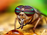 horsefly(female)
 -


_(1).jpg)

Siphoning
The
In species of Lepidoptera, it consists of two tubes held together by hooks and separable for cleaning. Each tube is inwardly concave, thus forming a central tube through which moisture is sucked. Suction is affected by the contraction and expansion of a sac in the head.
- Siphoning mouthparts
-
-

-

-


.jpg)
Sponging
The mouthparts of insects that feed on fluids are modified in various ways to form a tube through which liquid can be drawn into the mouth and usually another through which saliva passes. The muscles of the cibarium or pharynx are strongly developed to form a pump.
The mouthparts of
- Sponging mouthparts
-

-

-


.jpg)

Thorax
The insect thorax has three segments: the prothorax, mesothorax, and metathorax. The anterior segment, closest to the head, is the prothorax; its major features are the first pair of legs and the pronotum. The middle segment is the mesothorax; its major features are the second pair of legs and the anterior wings, if any. The third, the posterior, thoracic segment, abutting the abdomen, is the metathorax, which bears the third pair of legs and the posterior wings. Each segment is delineated by an intersegmental suture. Each segment has four basic regions. The dorsal surface is called the tergum (or notum, to distinguish it from the abdominal terga).[1]: 22–24 The two lateral regions are called the pleura (singular: pleuron), and the ventral aspect is called the sternum. In turn, the notum of the prothorax is called the pronotum, the notum for the mesothorax is called the mesonotum and the notum for the metathorax is called the metanotum. Continuing with this logic, there are also the mesopleura and metapleura, as well as the mesosternum and metasternum.[8]
The tergal plates of the thorax are simple structures in apterygotes and many immature insects but are variously modified in winged adults. The pterothoracic nota each have two main divisions: the anterior, wing-bearing alinotum and the posterior, phragma-bearing postnotum. Phragmata (singular: phragma) are plate-like apodemes that extend inwards below the antecostal sutures, marking the primary intersegmental folds between segments; phragmata provide attachment for the longitudinal flight muscles. Each alinotum (sometimes confusingly referred to as a "notum") may be traversed by sutures that mark the position of internal strengthening ridges and commonly divide the plate into three areas: the anterior prescutum, the scutum, and the smaller posterior scutellum. The lateral pleural sclerites are believed to be derived from the subcoxal segment of the ancestral insect leg. These sclerites may be separate, as in silverfish, or fused into an almost continuous sclerotic area, as in most winged insects.[1]: 22–24
Prothorax
The pronotum of the prothorax may be simple in structure and small in comparison with the other nota, but in beetles, mantids, many bugs, and some Orthoptera, the pronotum is expanded, and in cockroaches, it forms a shield that covers part of the head and mesothorax.[8][1]: 22–24
Pterothorax
Because the mesothorax and metathorax hold the wings, they have a combined name called the pterothorax (pteron = wing). The forewing, which goes by different names in different orders (e.g., the tegmina in Orthoptera and elytra in Coleoptera), arises between the mesonotum and the mesopleuron, and the hindwing articulates between the metanotum and metapleuron. The legs arise from the mesopleuron and metapleura. The mesothorax and metathorax each have a pleural suture (mesopleural and metapleural sutures) that runs from the wing base to the coxa of the leg. The sclerite anterior to the pleural suture is called the episternum (serially, the mesepisternum and metepisternum). The sclerite posterior to the suture is called the epimiron (serially, the mesepimiron and metepimiron). Spiracles, the external organs of the respiratory system, are found on the pterothorax, usually one between the pro- and mesopleoron, as well as one between the meso- and metapleuron.[8]
The ventral view or sternum follows the same convention, with the prosternum under the prothorax, the mesosternum under the mesothorax and the metasternum under the metathorax. The notum, pleura, and sternum of each segment have a variety of different sclerites and sutures, varying greatly from order to order, and they will not be discussed in detail in this section.[8]
Wings
Most phylogenetically advanced insects have two pairs of wings located on the second and third thoracic segments.[1]: 22–24 Insects are the only invertebrates to have developed flight capability, and this has played an important part in their success. Insect flight is not very well understood, relying on turbulent aerodynamic effects. The primitive insect groups use muscles that act directly on the wing structure. The more advanced groups making up the Neoptera have foldable wings, and their muscles act on the thorax wall and power the wings indirectly.[1]: 22–24 These muscles can contract multiple times for each single nerve impulse, allowing the wings to beat faster than would ordinarily be possible.
Insect flight can be rapid, maneuverable, and versatile, possibly due to the changing shape, extraordinary control, and variable motion of the insect wing. Insect orders use different flight mechanisms; for example, the flight of a butterfly can be explained using steady-state, nontransitory aerodynamics, and thin airfoil theory.

Internal
Each of the wings consists of a thin membrane supported by a system of veins. The membrane is formed by two layers of integument closely apposed, while the veins are formed where the two layers remain separate and the cuticle may be thicker and more heavily sclerotized. Within each of the major veins is a nerve and a trachea, and, since the cavities of the veins are connected with the
Veins

In some minuscule insects, the venation may be reduced. In
The archedictyon is the name given to a hypothetical scheme of wing venation proposed for the very first winged insect. It is based on a combination of speculation and fossil data. Since all winged insects are believed to have evolved from a common ancestor, the archediction represents the "template" that has been modified (and streamlined) by natural selection for 200 million years. According to current dogma, the archedictyon contained six to eight longitudinal veins. These veins (and their branches) are named according to a system devised by John Comstock and George Needham—the Comstock-Needham system:[37]
- Costa (C) – the leading edge of the wing
- Subcosta (Sc) – second longitudinal vein (behind the costa), typically unbranched
- Radius (R) – third longitudinal vein, one to five branches reach the wing margin
- Media (M) – fourth longitudinal vein, one to four branches reach the wing margin
- Cubitus (Cu) – fifth longitudinal vein, one to three branches reach the wing margin
- Anal veins (A1, A2, A3) – unbranched veins behind the cubitus
The costa (C) is the leading marginal vein on most insects, although a small vein, the precosta, is sometimes found above the costa. In almost all extant insects,[1]: 41–42 the precosta is fused with the costa; the costa rarely ever branches because it is at the leading edge, which is associated at its base with the humeral plate. The trachea of the costal vein is perhaps a branch of the subcostal trachea. Located after the costa is the third vein, the subcosta, which branches into two separate veins: the anterior and posterior. The base of the subcosta is associated with the distal end of the neck of the first axillary. The fourth vein is the radius, which is branched into five separate veins. The radius is generally the strongest vein of the wing. Toward the middle of the wing, it forks into a first undivided branch (R1) and a second branch, called the radial sector (Ra), which subdivides dichotomously into four distal branches (R2, R3, R4, R5). Basally, the radius is flexibly united with the anterior end of the second axillary (2Ax).[38]
The fifth vein of the wing is the media. In the archetype pattern (A), the media forks into two main branches, a media anterior (MA), which divides into two distal branches (MA1, MA2), and a median sector, or media posterior (MP), which has four terminal branches (M1, M2, M3, M4). In most modern insects, the media anterior has been lost, and the usual "media" is the four-branched media posterior with the common basal stem. In the Ephemerida, according to present interpretations of the wing venation, both branches of the media are retained, while in Odonata, the persisting media is the primitive anterior branch. The stem of the media is often united with the radius, but when it occurs as a distinct vein, its base is associated with the distal median plate (m') or is continuously sclerotized with the latter. The cubitus, the sixth vein of the wing, is primarily two-branched. The primary forking takes place near the base of the wing, forming the two principal branches (Cu1, Cu2). The anterior branch may break up into several secondary branches, but commonly it forks into two distal branches. The second branch of the cubitus (Cu2) in Hymenoptera, Trichoptera, and Lepidoptera, was mistaken by Comstock and Needham for the first anal. Proximally, the main stem of the cubitus is associated with the distal median plate (m') of the wing base.[38]
The postcubitus (Pcu) is the first anal of the Comstock and Needham system. The postcubitus, however, has the status of an independent wing vein and should be recognized as such. In nymphal wings, its trachea arises between the cubital trachea and the group of vannal tracheae. In the mature wings of more generalized insects, the postcubitus is always associated proximally with the cubitus and is never intimately connected with the flexor sclerite (3Ax) of the wing base. In Neuroptera, Mecoptera, and Trichoptera, the postcubitus may be more closely associated with the vannal veins, but its base is always free from the latter. The postcubitus is usually unbranched; primitively, it is two-branched. The vannal veins (lV to nV) are the anal veins immediately associated with the third axillary, and are directly affected by the movement of this sclerite that brings about the flexion of the wings. In number, the vannal veins vary from one to 12, according to the expansion of the vannal area of the wing. The vannal tracheae usually arise from a common tracheal stem in nymphal insects, and the veins are regarded as branches of a single anal vein. Distally, the vannal veins are either simple or branched. The jugal vein (J) of the jugal lobe of the wing is often occupied by a network of irregular veins, or it may be entirely membranous; sometimes it contains one or two distinct, small veins, the first jugal vein, or vena arcuata, and the second jugal vein, or vena cardinalis (2J).[38]
- C-Sc cross-veins – run between the costa and subcosta
- R cross-veins – run between adjacent branches of the radius
- R-M cross-veins – run between the radius and media
- M-Cu cross-veins – run between the media and cubitus
All the veins of the wing are subject to secondary forking and union by cross-veins. In some orders of insects, the cross-veins are so numerous, the whole venational pattern becomes a close network of branching veins and cross-veins. Ordinarily, however, a definite number of cross-veins having specific locations occurs. The more constant cross-veins are the humeral cross-vein (h) between the costa and subcosta, the radial cross-vein (r) between R and the first fork of Rs, the sectorial cross-vein (s) between the two forks of R8, the median cross-vein (m-m) between M2 and M3, and the mediocubital cross-vein (m-cu) between the media and the cubitus.[38]
The veins of insect wings are characterized by a convex-concave placement, such as those seen in mayflies (i.e., concave is "down" and convex is "up"), which alternate regularly and by their branching; whenever a vein forks there is always an interpolated vein of the opposite position between the two branches. The concave vein will fork into two concave veins (with the interpolated vein being convex) and the regular alteration of the veins is preserved.[39] The veins of the wing appear to fall into an undulating pattern according to whether they tend to fold up or down when the wing is relaxed. The basal shafts of the veins are convex, but each vein forks distally into an anterior convex branch and a posterior concave branch. Thus, the costa and subcosta are regarded as convex and concave branches of a primary first vein, Rs is the concave branch of the radius, posterior media is the concave branch of the media, Cu1 and Cu2 are respectively convex and concave, while the primitive postcubitus and the first vannal have each an anterior convex branch and a posterior concave branch. The convex or concave nature of the veins has been used as evidence in determining the identities of the persisting distal branches of the veins of modern insects, but it has not been demonstrated to be consistent for all wings.[26][38]
Fields

Wing areas are delimited and subdivided by
- Remigium
- Anal area (vannus)
- Jugal area
- Axillary area
- Alula
Most veins and cross-veins occur in the anterior area of the remigium, which is responsible for most of the flight, powered by the thoracic muscles. The posterior portion of the remigium is sometimes called the clavus; the two other posterior fields are the anal and jugal areas.[1]: 41–42 When the vannal fold has the usual position anterior to the group of anal veins, the remigium contains the costal, subcostal, radial, medial, cubital, and postcubital veins. In the flexed wing, the remigium turns posteriorly on the flexible basal connection of the radius with the second axillary, and the base of the mediocubital field is folded medially on the axillary region along the plica basalis (bf) between the median plates (m, m') of the wing base.[38]
The vannus is bordered by the vannal fold, which typically occurs between the postcubitus and the first vannal vein. In Orthoptera, it usually has this position. In the forewing of Blattidae, however, the only fold in this part of the wing lies immediately before the postcubitus. In Plecoptera, the vannal fold is posterior to the postcubitus, but proximally it crosses the base of the first vannal vein. In the cicada, the vannal fold lies immediately behind the first vannal vein (lV). These small variations in the actual position of the vannal fold, however, do not affect the unity of action of the vannal veins, controlled by the flexor sclerite (3Ax), in the flexion of the wing. In the hindwings of most Orthoptera, a secondary vena dividens forms a rib in the vannal fold. The vannus is usually triangular in shape, and its veins typically spread out from the third axillary like the ribs of a fan. Some of the vannal veins may be branched, and secondary veins may alternate with the primary veins. The vannal region is usually best developed in the hindwing, in which it may be enlarged to form a sustaining surface, as in Plecoptera and Orthoptera. The great fan-like expansions of the hindwings of Acrididae are clearly the vannal regions, since their veins are all supported on the third axillary sclerites on the wing bases, though Martynov (1925) ascribes most of the fan areas in Acrididae to the jugal regions of the wings. The true jugum of the acridid wing is represented only by the small membrane (Ju) mesad of the last vannal vein. The jugum is more highly developed in some other Orthoptera, as in the Mantidae. In most of the higher insects with narrow wings, the vannus becomes reduced, and the vannal fold is lost, but even in such cases, the flexed wing may bend along a line between the postcubitus and the first vannal vein.[38]
The jugal region, or neala, is a region of the wing that is usually a small membranous area proximal to the base of the vannus strengthened by a few small, irregular vein-like thickenings; but when well developed, it is a distinct section of the wing and may contain one or two jugal veins. When the jugal area of the forewing is developed as a free lobe, it projects beneath the humeral angle of the hindwing and thus serves to yoke the two wings together. In the Jugatae group of Lepidoptera, it bears a long finger-like lobe. The jugal region was termed the neala ("new wing") because it is a secondary and recently developed part of the wing.[38]
The auxiliary region containing the axillary sclerites has, in general, the form of a scalene triangle. The base of the triangle (a-b) is the hinge of the wing with the body; the apex (c) is the distal end of the third axillary sclerite; the longer side is anterior to the apex. Point d on the anterior side of the triangle marks the articulation of the radial vein with the second axillary sclerite. The line between d and c is the plica basalis (bf), or fold of the wing at the base of the mediocubital field.[38]
At the posterior angle of the wing base in some Diptera, there is a pair of membranous lobes (squamae, or calypteres) known as the alula. The alula is well developed in the house fly. The outer squama (c) arises from the wing base behind the third axillary sclerite (3Ax) and represents the jugal lobe of other insects (A, D); the larger inner squama (d) arises from the posterior scutellar margin of the tergum of the wing-bearing segment and forms a protective, hood-like canopy over the halter. In the flexed wing, the outer squama of the alula is turned upside down above the inner squama, the latter not being affected by the movement of the wing. In many Diptera, a deep incision of the anal area of the wing membrane behind the single vannal vein sets off a proximal alar lobe distal to the outer squama of the alula.[38]
Joints

The various movements of the wings, especially in insects that flex their wings horizontally over their backs when at rest, demand a more complicated articular structure at the wing base than a mere hinge of the wing with the body. Each wing is attached to the body by a membranous basal area, but the articular membrane contain several small articular sclerites, collectively known as the pteralia. The pteralia include an anterior humeral plate at the base of the costal vein, a group of axillaries (Ax) associated with the subcostal, radial, and vannal veins, and two less definite median plates (m, m') at the base of the mediocubital area. The axillaries are specifically developed only in wing-flexing insects, where they constitute the flexor mechanism of the wing operated by the flexor muscle arising on the pleuron. Characteristic of the wing base is also a small lobe on the anterior margin of the articular area proximal to the humeral plate, which, in the forewing of some insects, is developed into a large, flat, scale-like flap, the tegula, overlapping the base of the wing. Posteriorly, the articular membrane often forms an ample lobe between the wing and the body, and its margin is generally thickened and corrugated, giving the appearance of a ligament, the so-called axillary cord, continuous mesally with the posterior marginal scutellar fold of the tergal plate bearing the wing.[38]
The articular sclerites, or pteralia, of the wing base of the wing-flexing insects and their relations to the body and the wing veins, shown diagrammatically, are as follows:
- Humeral plates
- First Axillary
- Second Axillary
- Third Axillary
- Fourth Axillary
- Median plates (m, m')
The humeral plate is usually a small sclerite on the anterior margin of the wing base, movable and articulated with the base of the costal vein. Odonata have their humeral plates greatly enlargened,[38] with two muscles arising from the episternum inserted into the humeral plates and two from the edge of the epimeron inserted into the axillary plate.[26]
The first axillary sclerite (lAx) is the anterior hinge plate of the wing base. Its anterior part is supported on the anterior notal wing process of the tergum (ANP); its posterior part articulates with the tergal margin. The anterior end of the sclerite is generally produced as a slender arm, the apex of which (e) is always associated with the base of the subcostal vein (Sc), though it is not united with the latter. The body of the sclerite articulates laterally with the second axillary. The second axillary sclerite (2Ax) is more variable in form than the first axillary, but its mechanical relations are no less definite. It is obliquely hinged to the outer margin of the body of the first axillary, and the radial vein (R) is always flexibly attached to its anterior end (d). The second axillary presents both a dorsal and ventral sclerotization in the wing base; its ventral surface rests upon the fulcral wing process of the pleuron. The second axillary, therefore, is the pivotal sclerite of the wing base, and it specifically manipulates the radial vein.[38]
The third axillary sclerite (3Ax) lies in the posterior part of the articular region of the wing. Its form is highly variable and often irregular, but the third axillary is the sclerite on which is inserted the flexor muscle of the wing (D). Mesally, it articulates anteriorly (f) with the posterior end of the second axillary, and posteriorly (b) with the posterior wing process of the tergum (PNP), or with a small fourth axillary when the latter is present. Distally, the third axillary is prolonged in a process always associated with the bases of the group of veins in the anal region of the wing, here termed the vannal veins (V). The third axillary, therefore, is usually the posterior hinge plate of the wing base and is the active sclerite of the flexor mechanism, which directly manipulates the vannal veins. The contraction of the flexor muscle (D) revolves the third axillary on its mesal articulations (b, f), and thereby lifts its distal arm; this movement produces the flexion of the wing. The fourth axillary sclerite is not a constant element of the wing base. When present, it is usually a small plate intervening between the third axillary and the posterior notal wing process and is probably a detached piece of the latter.[38]
The median plates (m, m') are also sclerites that are not so definitely differentiated as specific plates as are the three principal axillaries, but they are important elements of the flexor apparatus. They lie in the median area of the wing base distal to the second and third axillaries and are separated from each other by an oblique line (bf), which forms a prominent convex fold during flexion of the wing. The proximal plate (m) is usually attached to the distal arm of the third axillary and perhaps should be regarded as a part of the latter. The distal plate (m') is less constantly present as a distinct sclerite and may be represented by a general sclerotization of the base of the mediocubital field of the wing. When the veins of this region are distinct at their bases, they are associated with the outer median plate.[38]
Coupling, folding, and other features
In many insect species, the forewing and hindwing are coupled together, which improves the aerodynamic efficiency of flight. The most common coupling mechanism (e.g.,
When at rest, the wings are held over the back in most insects, which may involve longitudinal folding of the wing membrane and sometimes also transverse folding. Folding may sometimes occur along the flexion lines. Though fold lines may be transverse, as in the hindwings of beetles and earwigs, they are normally radial to the base of the wing, allowing adjacent sections of a wing to be folded over or under each other. The commonest fold line is the jugal fold, situated just behind the third anal vein,[27] although, most Neoptera have a jugal fold just behind vein 3A on the forewings. It is sometimes also present on the hindwings. Where the anal area of the hindwing is large, as in Orthoptera and Blattodea, the whole of this part may be folded under the anterior part of the wing along a vannal fold a little posterior to the claval furrow. In addition, in Orthoptera and Blattodea, the anal area is folded like a fan along the veins, the anal veins being convex, at the crests of the folds, and the accessory veins concave. Whereas the claval furrow and jugal fold are probably homologous in different species, the vannal fold varies in position in different taxa. Folding is produced by a muscle arising on the pleuron and inserted into the third axillary sclerite in such a way that when it contracts, the sclerite pivots about its points of articulation with the posterior notal process and the second axillary sclerite.[26]
As a result, the distal arm of the third axillary sclerite rotates upwards and inwards, so that finally its position is completely reversed. The anal veins are articulated with this sclerite in such a way that when it moves they are carried with it and become flexed over the back of the insect. Activity of the same muscle in flight affects the power output of the wing and so it is also important in flight control. In orthopteroid insects, the elasticity of the cuticle causes the vannal area of the wing to fold along the veins. Consequently, energy is expended in unfolding this region when the wings are moved to the flight position. In general, wing extension probably results from the contraction of muscles attached to the basilar sclerite or, in some insects, to the subalar sclerite.[26]
Legs

The typical and usual segments of the
The inflection of the coxal wall bearing the pleural articular surface divides the lateral wall of the basicoxite into a prearticular part and a postarticular part, and the two areas often appear as two marginal lobes on the base of the coxa. The posterior lobe is usually the larger and is termed the meron. The meron may be greatly enlarged by an extension distally in the posterior wall of the coxa; in the Neuroptera, Mecoptera, Trichoptera, and Lepidoptera, the meron is so large that the coxa appears to be divided into an anterior piece, the so-called "coxa genuina," and the meron, but the meron never includes the region of the posterior trochanteral articulation, and the groove delimiting it is always a part of the basicostal suture. A coxa with an enlarged meron has an appearance similar to one divided by a coxal suture falling in line with the pleural suture, but the two conditions are fundamentally quite different and should not be confused. The meron reaches the extreme of its departure from the usual condition in the Diptera. In some of the more generalized flies, as in the Tipulidae, the meron of the middle leg appears as a large lobe of the coxa projecting upward and posteriorly from the coxal base; in higher members of the order, it becomes completely separated from the coxa and forms a plate of the lateral wall of the mesothorax.[38]: 164
The trochanter is the basal segment of the telopodite; it is always a small segment in the insect leg, freely movable by a horizontal hinge on the coxa, but more or less fixed to the base of the femur. When movable on the femur the trochantero femoral hinge is usually vertical or oblique in a vertical plane, giving a slight movement of production and reduction at the joint, though only a reductor muscle is present. In the Odonata, both nymphs and adults, there are two trochanteral segments, but they are not movable on each other; the second contains the reductor muscle of the femur. The usual single trochanteral segment of insects, therefore, probably represents the two trochanters of other arthropods fused into one apparent segment since it is not likely that the primary coxotrochanteral hinge has been lost from the leg. In some of the Hymenoptera, a basal subdivision of the femur simulates a second trochanter, but the insertion of the reductor muscle on its base attests that it belongs to the femoral segment, since as shown in the odonate leg, the reductor has its origin in the true second trochanter.[38]: 165
The femur is the third segment of the insect leg, is usually the longest and strongest part of the limb, but it varies in size from the huge hind femur of leaping Orthoptera to a very small segment such as is present in many larval forms. The volume of the femur is generally correlated with the size of the tibial muscles contained within it, but it is sometimes enlarged and modified in shape for other purposes than that of accommodating the tibial muscles. The tibia is characteristically a slender segment in adult insects, only a little shorter than the femur or the combined femur and trochanter. Its proximal end forms a more or less distinct head bent toward the femur, a device allowing the tibia to be flexed close against the undersurface of the femur.[38]: 165
The terms profemur, mesofemur, and metafemur refer to the femora of the front, middle and hind legs of an insect, respectively.[40] Similarly, protibia, mesotibia, and metatibia refer to the tibiae of the front, middle and hind legs.[41]
The tarsus of insects corresponds to the penultimate segment of a generalized arthropod limb, which is the segment called the propodite in Crustacea. In adult insects, it is commonly subdivided into two to five subsegments, or tarsomeres, but in the Protura, some Collembola, and most holometabolous insect larvae it preserves the primitive form of a simple segment. The subsegments of the adult insect tarsus are usually freely movable on one another by inflected connecting membranes, but the tarsus never has intrinsic muscles. The tarsus of adult pterygote insects having fewer than five subsegments is probably specialized by the loss of one or more subsegments or by a fusion of adjoining subsegments. In the tarsi of Acrididae, the long basal piece is composed of three united tarsomeres, leaving the fourth and the fifth. The basal tarsomere is sometimes conspicuously enlarged and is distinguished as the basitarsus. On the under surfaces of the tarsal subsegments in certain Orthoptera, there are small pads, the tarsal pulvilli, or euplantulae. The tarsus is occasionally fused with the tibia in larval insects, forming a tibiotarsal segment; in some cases, it appears to be eliminated or reduced to a rudiment between the tibia and the pretarsus.[38]: 165–166
For the most part, the femur and tibia are the longest leg segments but variations in the lengths and robustness of each segment relate to their functions. For example, gressorial and cursorial, or walking and running type insects respectively, usually have well-developed
Abdomen
The ground plan of the abdomen of an adult insect typically consists of 11–12 segments and is less strongly sclerotized than the head or thorax. Each segment of the abdomen is represented by a sclerotized tergum, sternum, and perhaps a pleurite. Terga are separated from each other and from the adjacent sterna or pleura by a membrane. Spiracles are located in the pleural area. Variation of this ground plan includes the fusion of terga or terga and sterna to form continuous dorsal or ventral shields or a conical tube. Some insects bear a sclerite in the pleural area called a laterotergite. Ventral sclerites are sometimes called laterosternites. During the embryonic stage of many insects and the postembryonic stage of primitive insects, 11 abdominal segments are present. In modern insects there is a tendency toward reduction in the number of the abdominal segments, but the primitive number of 11 is maintained during embryogenesis. Variation in abdominal segment number is considerable. If the Apterygota are considered to be indicative of the ground plan for pterygotes, confusion reigns: adult Protura have 12 segments, Collembola have 6. The orthopteran family Acrididae has 11 segments, and a fossil specimen of Zoraptera has a 10-segmented abdomen.[8]
Generally, the first seven abdominal segments of adults (the pregenital segments) are similar in structure and lack appendages. However, apterygotes (bristletails and silverfish) and many immature aquatic insects have abdominal appendages. Apterygotes possess a pair of styles; rudimentary appendages that are serially homologous with the distal part of the thoracic legs. And, mesally, one or two pairs of protrusible (or exsertile) vesicles on at least some abdominal segments. These vesicles are derived from the coxal and trochanteral endites (inner annulated lobes) of the ancestral abdominal appendages. Aquatic larvae and nymphs may have gills laterally on some to most abdominal segments.[1]: 49 Of the rest of the abdominal segments consist of the reproductive and anal parts.
External genitalia
The organs concerned specifically with mating and the deposition of eggs are known collectively as the external genitalia, although they may be largely internal. The components of the external genitalia of insects are very diverse in form and often have considerable taxonomic value, particularly among species that appear structurally similar in other respects. The male external genitalia have been used widely to aid in distinguishing species, whereas the female external genitalia may be simpler and less varied.
The terminalia of adult female insects include internal structures for receiving the male copulatory organ and his spermatozoa and external structures used for oviposition (egg-laying; section 5.8). Segments 8 and 9 bear the genitalia; segment 10 is visible as a complete segment in many "lower" insects but always lacks appendages. Most female insects have an egg-laying tube, or ovipositor; it is absent in termites, parasitic lice, many Plecoptera, and most Ephemeroptera. Ovipositors take two forms:
- The anal-genital part of the abdomen. which consists generally of segments 8 or 9 to the abdominal apex
- substitutional, composed of extensible posterior abdominal segments.
Other appendages
The terminal abdominal segments have excretory and sensory functions in all insects, besides the reproductive function in adults.
- the paraprocts: paired plate-like appendages also derived from the sternum at the side of the tip of the abdomen, often most apparent in certain basal orders such as Odonata;
- the cerci: a pair of appendages which articulate laterally on segment 11; typically, these are annulated and filamentous but have been modified (e.g. the forceps of earwigs) or reduced in different insect orders.
- a central caudal filament, prolongation or median appendix dorsalis, which arises from the tip of the epiproct in certain Ephemeroptera), and a few fossil insects.[42] A similar structure in nymphal stoneflies (Plecoptera) is of uncertain homology.
Internal
Nervous system
The
The thoracic segments have one ganglion on each side, which are connected into a pair, one pair per segment. This arrangement is also seen in the abdomen but only in the first eight segments. Many species of insects have reduced numbers of ganglia due to fusion or reduction.
At least a few insects have
Digestive system
An insect uses its digestive system for all steps in food processing: digestion, absorption, and feces delivery and elimination.
Foregut

The first section of the alimentary canal is the
From there, the pharynx passes food to the esophagus, which could be just a simple tube passing it on to the crop and proventriculus, and then on ward to the midgut, as in most insects. Alternately, the foregut may expand into a very enlarged crop and proventriculus, or the crop could just be a diverticulum, or fluid filled structure, as in some Diptera species.[50]: 30–31

The
Midgut
Once food leaves the crop, it passes to the midgut (element 13 in numbered diagram), also known as the mesenteron, where the majority of digestion takes place. Microscopic projections from the midgut wall, called microvilli, increase the surface area of the wall and allow more nutrients to be absorbed; they tend to be close to the origin of the midgut. In some insects, the role of the microvilli and where they are located may vary. For example, specialized microvilli producing digestive enzymes may more likely be near the end of the midgut, and absorption near the origin or beginning of the midgut.[50]: 32
In the wingless (apterygote) orders Archaeognatha and Zygentoma (and the hexapods Entognatha), the midgut epithelium is derived entirely from yolk cells. In the majority of the flying insects (Neoptera), it is derived from bipolar formation. The Palaeoptera (mayflies and dragonflies) show a transition between apterygotes and neopterans, where the middle part of the midgut epithelium is derived from yolk cells and the anterior and posterior parts are formed through bipolar formation.[52]
Hindgut
In the hindgut (element 16 in numbered diagram), or proctodaeum, undigested food particles are joined by uric acid to form fecal pellets. The rectum absorbs 90% of the water in these fecal pellets, and the dry pellet is then eliminated through the anus (element 17), completing the process of digestion. The uric acid is formed using hemolymph waste products diffused from the Malpighian tubules (element 20). It is then emptied directly into the alimentary canal, at the junction between the midgut and hindgut. The number of Malpighian tubules possessed by a given insect varies between species, ranging from only two tubules in some insects to over 100 tubules in others.[1]: 71–72, 78–80
Respiratory systems
There are many different patterns of gas exchange demonstrated by different groups of insects. Gas exchange patterns in insects can range from continuous and diffusive ventilation, to discontinuous gas exchange.[1]: 65–68 During continuous gas exchange, oxygen is taken in and carbon dioxide is released in a continuous cycle. In discontinuous gas exchange, however, the insect takes in oxygen while it is active and small amounts of carbon dioxide are released when the insect is at rest.[54] Diffusive ventilation is simply a form of continuous gas exchange that occurs by diffusion rather than physically taking in the oxygen. Some species of insect that are submerged also have adaptations to aid in respiration. As larvae, many insects have gills that can extract oxygen dissolved in water, while others need to rise to the water surface to replenish air supplies, which may be held or trapped in special structures.[55][56]
Circulatory system
Insect blood or haemolymph's main function is that of transport and it bathes the insect's body organs. Making up usually less than 25% of an insect's body weight, it transports
Body fluids enter through one-way valved ostia, which are openings situated along the length of the combined aorta and heart organ. Pumping of the haemolymph occurs by waves of peristaltic contraction, originating at the body's posterior end, pumping forwards into the dorsal vessel, out via the aorta and then into the head where it flows out into the haemocoel.
Endocrine system
These glands are part of the endocrine system:
1. Neurosecretory cells
2. Corpora cardiaca
Reproductive system
Female
Female insects are able make eggs, receive and store sperm, manipulate sperm from different males, and lay eggs. Their reproductive systems are made up of a pair of
The ovaries are made up of a number of egg tubes, called
Accessory glands or glandular parts of the oviducts produce a variety of substances for sperm maintenance, transport, and fertilization, as well as for protection of eggs. They can produce glue and protective substances for coating eggs or tough coverings for a batch of eggs called oothecae. Spermathecae are tubes or sacs in which sperm can be stored between the time of mating and the time an egg is fertilized. Paternity testing of insects has revealed that some, and probably many, female insects use the spermatheca and various ducts to control or bias sperm used in favor of some males over others.[8]: 880
Male
The main component of the male reproductive system is the
Internal morphology of different taxa
Blattodea
Cockroaches are most common in tropical and
Cockroaches, like all insects, breathe through a system of tubes called
Coleoptera
The
The
Like other insect species, beetles have
Different glands specialize for different pheromones produced for finding mates. Pheromones from species of
A notable number of species have developed special glands that produce chemicals for deterring predators (see Defense and predation). The Ground beetle's (of Carabidae) defensive glands, located at the posterior, produce a variety of hydrocarbons, aldehydes, phenols, quinones, esters, and acids released from an opening at the end of the abdomen. While African carabid beetles (e.g., Anthia some of which used to comprise the genus Thermophilum) employ the same chemicals as ants: formic acid.[31] While Bombardier beetles have well developed, like other carabid beetles, pygidial glands that empty from the lateral edges of the intersegment membranes between the seventh and eighth abdominal segments. The gland is made of two containing chambers. The first holds hydroquinones and hydrogen peroxide, with the second holding just hydrogen peroxide plus catalases. These chemicals mix and result in an explosive ejection, forming temperatures of around 100 C, with the breakdown of hydroquinone to H2 + O2 + quinone, with the O2 propelling the excretion.[8]
Tympanal organs are hearing organs. Such an organ is generally a membrane (tympanum) stretched across a frame backed by an air sac and associated sensory neurones. In the order Coleoptera, tympanal organs have been described in at least two families.[30] Several species of the genus Cicindela in the family Cicindelidae have ears on the dorsal surface of the first abdominal segment beneath the wing; two tribes in the family Dynastinae (Scarabaeidae) have ears just beneath the pronotal shield or neck membrane. The ears of both families are to ultrasonic frequencies, with strong evidence that they function to detect the presence of bats via their ultrasonic echolocation. Even though beetles constitute a large order and live in a variety of niches, examples of hearing is surprisingly lacking in species, though it is likely that most are just undiscovered.[8]
Dermaptera
The
The reproductive system of females consist of paired ovaries, lateral oviducts, spermatheca, and a genital chamber. The lateral ducts are where the eggs leave the body, while the spermatheca is where sperm is stored. Unlike other insects, the gonopore, or genital opening is behind the seventh abdominal segment. The ovaries are primitive in that they are polytrophic (the nurse cells and oocytes alternate along the length of the ovariole). In some species these long ovarioles branch off the lateral duct, while in others, short ovarioles appear around the duct.[8]
Diptera
The genitalia of female flies are rotated to a varying degree from the position found in other insects. In some flies this is a temporary rotation during mating, but in others it is a permanent torsion of the organs that occurs during the pupal stage. This torsion may lead to the anus being located below the genitals, or, in the case of 360° torsion, to the sperm duct being wrapped around the gut, despite the external organs being in their usual position. When flies mate, the male initially flies on top of the female, facing in the same direction, but then turns round to face in the opposite direction. This forces the male to lie on its back in order for its genitalia to remain engaged with those of the female, or the torsion of the male genitals allows the male to mate while remaining upright. This leads to flies having more reproduction abilities than most insects and at a much quicker rate. Flies come in great populations due to their ability to mate effectively and in a short period of time especially during the mating season.[71]
The female lays her eggs as close to the food source as possible, and development is very rapid, allowing the larva to consume as much food as possible in a short period of time before transforming into the adult. The eggs hatch soon after being laid, or the flies are
take various forms, and in some cases develop inside a silk cocoon. After emerging from the pupa, the adult fly rarely lives more than a few days, and serves mainly to reproduce and to disperse in search of new food sources.Lepidoptera
In
In the digestive system, the anterior region of the foregut has been modified to form a pharyngeal sucking pump as they need it for the food they eat, which are for the most part liquids. An esophagus follows and leads to the posterior of the pharynx and in some species forms a form of crop. The midgut is short and straight, with the hindgut being longer and coiled.[69] Ancestors of lepidopteran species, stemming from Hymenoptera, had midgut ceca, although this is lost in current butterflies and moths. Instead, all the digestive enzymes other than initial digestion, are immobilized at the surface of the midgut cells. In larvae, long-necked and stalked goblet cells are found in the anterior and posterior midgut regions, respectively. In insects, the goblet cells excrete positive potassium ions, which are absorbed from leaves ingested by the larvae. Most butterflies and moths display the usual digestive cycle, however species that have a different diet require adaptations to meet these new demands.[8]: 279
In the
See also
- Morphology (biology)
- Insect physiology
- External morphology of Lepidoptera
- External morphology of Odonata
- Insect ecology
- Insect flight
- Invertebrate
- Insect
- Entomology
- Prehistoric insect
References
- ^ ISBN 1-4051-1113-5.
- ^ "O. Orkin Insect zoo". The University of Nebraska Department of Entomology. Archived from the original on 2009-06-02. Retrieved 2009-05-03.
- ^ Resh, Vincent H.; Cardé, Ring T. (2009). Encyclopedia of Insects (2nd ed.). San Diego, CA: Academic Press. p. 12.
- ISBN 0-8053-1957-3
- ISBN 978-0-595-22143-1.
- ^ a b "external morphology of Insects" (PDF). Archived from the original (PDF) on 2011-07-19. Retrieved 2011-03-20.
- ^ ISBN 0-412-61390-5.
- ^ ISBN 978-0-12-374144-8.
- ^ Smith, John Bernhard, Explanation of terms used in entomology Publisher: Brooklyn entomological society 1906 (May be downloaded from: https://archive.org/details/explanationofter00smit)
- ^ Fox, Richard (6 Oct 2006). "External Anatomy". Lander University. Archived from the original on 2011-03-14. Retrieved 2011-03-20.
- ^ Volkel, R.; Eisner, M.; Weible, K.J. "Miniaturized imaging systems" (PDF). SUSS MicroOptics. Archived from the original (PDF) on 2008-10-01. Retrieved 2011-03-20.
{{cite journal}}: Cite journal requires|journal=(help) - PMID 4854430.
- ISSN 0167-9317.
- .
- .
- PMID 18089073.
- ^ ISBN 0-521-57048-4.
- S2CID 11851224.
- PMID 11815654.
- ISBN 978-0-12-024232-0.
- ^ Servadei, A.; Zangheri, S.; Masutti, L. (1972). Entomologia generale ed applicata. CEDAM. pp. 492–530.
- .
- ^ "Insect antennae". The Amateur Entomologists' Society. Retrieved 2011-03-21.
- ^ a b "Insect Morphology". University of Minisotta (Department of Entomology). Archived from the original on 2011-03-03. Retrieved 2011-03-21.
- ^ Kirejtshuk, A.G. (November 2002). "Head". Beetles (Coleoptera) and coleopterologist. zin.ru. Retrieved 2011-03-21.
- ^ ISBN 0-521-57048-4.
- ^ ISBN 0-306-44967-6.
- ISBN 1-57808-024-X.
- ^ "Mosquito biting mouthparts". allmosquitos.com. 2011. Retrieved April 17, 2011.
- ^ ISBN 978-1-4020-6242-1.
- ^ ISBN 0-520-22323-3.
- ISBN 978-1-4020-6242-1.
- .
- .
- ^ "Sponging". University of Minissota. Archived from the original on September 27, 2011. Retrieved April 17, 2011.
- ^ "Fly Mouthparts". School of Biological Sciences Online Learning Resources. University of Sydney. February 4, 2010. Archived from the original on April 9, 2011. Retrieved April 17, 2011.
- ^ Meyer, John R. (5 January 2007). "External Anatomy: WINGS". Department of Entomology, NC State University. Archived from the original on 16 July 2011. Retrieved 2011-03-21.
- ^ ISBN 0-8014-8125-2.
- ^ Spieth, HT (1932). "A New Method of Studying the Wing Veins of the Mayflies and Some Results Therefrom (Ephemerida)" (PDF). Entomological News. Archived from the original (PDF) on 2011-09-30.
- ^ "profemur, profemora – BugGuide.Net". bugguide.net. Retrieved 10 December 2016.
- ^ "protibia – BugGuide.Net". bugguide.net. Retrieved 10 December 2016.
- ^ Amateur Entomologist's Society: epiproct definition
- JSTOR 1539265.
- S2CID 3071.
- S2CID 1424315.
- ^ Sømme, LS (14 January 2005). "Sentience and pain in invertebrates". Norwegian Scientific Committee for Food Safety. Retrieved September 30, 2009.[permanent dead link]
- ^ a b "General Entomology – Digestive and Excritory system". NC state University. Retrieved 2009-05-03.
- ISBN 978-0-12-374144-8, retrieved 2020-09-29
- ISBN 978-0-12-374144-8, retrieved 2020-09-29
- ^ ISBN 0-8493-1181-0.
- ^ Duncan, Carl D. (1939). A Contribution to The Biology of North American Vespine Wasps (1 ed.). Stanford: Stanford University Press. pp. 24–29.
- ^ Revisiting the formation of midgut epithelium in Zygentoma (Insecta) from a developmental study of the firebrat Thermobia domestica (Packard, 1873) (Lepismatidae)
- ^ Meyer, John R. (17 February 2006). "Circulatory System". NC State University: Department of Entomology, NC State University. p. 1. Archived from the original on 27 September 2009. Retrieved 2009-10-11.
- ISBN 0-19-851549-9.
- ISBN 978-0-7575-5049-2.)
{{cite book}}:|author=has generic name (help)CS1 maint: multiple names: authors list (link - ISBN 978-0-7575-4128-5.)
{{cite book}}: CS1 maint: multiple names: authors list (link - ^ ISBN 978-0-19-850002-5.
- ^ a b c Triplehorn, C. A.; Johnson, N. F. (2005). Borror and DeLong's Introduction to the Study of Insects. Brooks / Thomson Cole: Brooks / Thomson Cole.
- ^ a b c Elzinga, R.J. (2004). Fundamentals of Entomology (6th ed.). New Jersey USA: Pearson/Prentice Hall.
- ISBN 978-0-03-096835-8.
- ISBN 1-4051-1113-5.
- ^ De Carlo; J. A. (1983). "Hemipteros acuáticos y semiacuáticos. Estudio en grupos en las partes de igual función de los aparatos genitales masculinos de especies estudiadas". Revista de la Sociedad Entomológica Argentina. 42 (1/4): 149–154.
- .
- S2CID 20011989.
- PMID 12716997.
- ^ Leung, Chee Chee Leung (March 22, 2007). "Cave may hold missing link". theage.com.au. Retrieved 7 December 2013.
- PMID 17376757.
- ^ Kunkel, Joseph G. "How do cockroaches breathe?". The Cockroach FAQ. UMass Amherst. Retrieved 7 December 2013.
- ^ ISBN 978-0-306-44967-3. Retrieved 14 November 2010.
- ISBN 0-521-57098-0. Retrieved 6 March 2010.
- ^ ISBN 0-19-510033-6.)
{{cite book}}: CS1 maint: multiple names: authors list (link - PMID 20268135.
- .
External links
- Insect Morphology Overview of insect external and internal anatomy
Anatomy and morphology | ||
|---|---|---|
| Fields |
|  |
| Bacteria and fungi | ||
| Protists | ||
| Plants |
| |
| Invertebrates |
| |
Mammals | ||
Other vertebrates |
| |
| Glossaries | ||
| Related topics | ||