
Intervertebral disc
| Intervertebral disc | |
|---|---|
spinal vertebrae | |
| Identifiers | |
| Latin | discus intervertebralis |
| MeSH | D007403 |
| TA98 | A03.2.02.003 |
| TA2 | 1684 |
| FMA | 10446 |
| Anatomical terminology] | |
An intervertebral disc (or intervertebral fibrocartilage) lies between adjacent
Structure

Intervertebral discs consist of an outer fibrous ring, the anulus (or annulus) fibrosus disci intervertebralis, which surrounds an inner gel-like center, the nucleus pulposus.
There is one disc between each pair of vertebrae, except for the first cervical segment, the
Development
During development and at birth, vertebral discs have some vascular supply to the cartilage endplates and the anulus fibrosus. These quickly deteriorate leaving almost no direct blood supply in healthy adults.[5]
Intervertebral disc space
The intervertebral disc space is typically defined on an
Function
The intervertebral disc functions to separate the vertebrae from each other and provides the surface for the shock-absorbing gel of the nucleus pulposus. The nucleus pulposus of the disc functions to distribute hydraulic pressure in all directions within each intervertebral disc under compressive loads. The nucleus pulposus consists of large
Clinical significance
Anything arising from the intervertebral disc may be termed discogenic in particular when referring to associated pain as discogenic pain.[7]
Herniation

A spinal disc herniation, commonly referred to as a slipped disc, can happen when unbalanced mechanical pressures substantially deform the anulus fibrosus, allowing part of the nucleus to obtrude. These events can occur during peak physical performance, during traumas, or as a result of chronic deterioration (typically accompanied with poor posture), and has been associated with a Propionibacterium acnes infection.[8] Both the deformed anulus and the gel-like material of the nucleus pulposus can be forced laterally or posteriorly, distorting local muscle function and putting pressure on the nearby nerve. This can give symptoms typical of nerve root entrapment, which can vary between paresthesia, numbness, chronic and/or acute pain, either locally or along the dermatome served by the entrapped nerve, loss of muscle tone and decreased homeostatic performance. The disc is not physically slipped; it bulges, usually in just one direction.
Another kind of herniation, of the nucleus pulposus, can happen as a result of the formation of Schmorl's nodes on the intervertebral disc.[9] This is referred to as vertical disc herniation.
Degeneration

Before age 40, approximately 25% of people show evidence of disc degeneration at one or more levels. Beyond age 40, more than 60% of people show evidence of disc degeneration at one or more levels on magnetic resonance imaging (MRI).[10] These degenerative changes are a normal part of the ageing process and do not correlate to pain.[11]

One effect of aging and disc degeneration is that the nucleus pulposus begins to dehydrate and the concentration of proteoglycans in the matrix decreases, thus limiting the ability of the disc to absorb shock. This general shrinking of disc size is partially responsible for the common decrease in height as humans age. The anulus fibrosus also becomes weaker with age and has an increased risk of tearing. In addition, the cartilage endplates begin thinning, fissures begin to form, and there is
Scoliosis
While this may not cause pain in some people, in others it may cause chronic pain. Other spinal disorders can affect the morphology of intervertebral discs. For example, patients with scoliosis commonly have calcium deposits (ectopic calcification) in the cartilage endplate and sometimes in the disc itself.[16] Herniated discs are also found to have a higher degree of cellular senescence than non-herniated discs.[5] In addition to scoliosis, which is the lateral 'S' curvature of the spine, the fused vertebrae can also experience other abnormalities such as kyphosis[12] (hunchback) which shows in old age, or lordosis[12] (swayback), which is often present in pregnancy and obesity.
Etymology
The Latin word anulus means "little ring"; it is the diminutive of anus ("ring"). However, modern English also spells the word more phonetically annulus, as with the term
Additional images
-
 Anulus fibrosus
 -
 Artificial intervertebral disc
 -
 Posterior longitudinal ligament, in the thoracic region
 -
 Median sagittal section through the occipital bone and first three cervical vertebrae
 -
 Costovertebral articulations. Anterior view.
 -
 Lumbar and sacral plexus. Deep dissection. Anterior view.
 -
 Lumbar and sacral plexus. Deep dissection. Anterior view.
 -
 Polarised light microscopy image of the annulus fibrosus, showing the concentric layers of fibrous tissue


.jpg)
See also
- Back pain
- Degenerative disc disease
- Spinal decompression
- Lumbar spinal stenosis
- List of distinct cell types in the adult human body
- Spelling of disc
References
- PMID 12723977.
- PMID 22028328.
- ^ "Intervertebral disc". Physio-pedia.com.
- PMID 24768732.
- ^ PMID 16595436.
- PMID 8770872.
- PMID 30270809.
- PMID 28369127.
- PMID 17530687.
- ^ "Intervertebral Disc Disorders". MDGuidelines. Reed Group. 1 December 2012. Archived from the original on 21 October 2012. Retrieved 1 December 2012.
- S2CID 33787081. Archived from the original(PDF) on 13 June 2020.
- ^ a b c "McGraw-Hill Connect". connect.mheducation.com. Retrieved 29 November 2015.
- ^ "Diagnosis & Treatment of Low Back Pain" (PDF). Spine.org. Retrieved 30 June 2022.
- PMID 415581.
- PMID 32084224.
- PMID 10079244.
- ^ Federative Committee on Anatomical Terminology (FCAT) (1998). Terminologia Anatomica. Stuttgart: Thieme
External links
- Intervertebral Discs
- Spinal Disc Summary
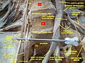
- Cross section image: pembody/body12a—Plastination Laboratory at the Medical University of Vienna
| National | |
|---|---|
| Other | |