
Intestinal villus
| Intestinal villus | |
|---|---|
mucosa showing villi – top half of image. H&E stain | |
 Section of duodenum of a cat. X 60. | |
| Details | |
| Part of | Wall of small intestine |
| System | Digestive system |
| Identifiers | |
| Latin | villi intestinales |
| TA98 | A05.6.01.011 |
| TA2 | 2941 |
| FMA | 15072 76464, 15072 |
| Anatomical terminology] | |
Intestinal villi (sg.: villus) are small, finger-like projections that extend into the
Villi increase the internal surface area of the intestinal walls making available a greater surface area for absorption. An increased absorptive area is useful because digested nutrients (including monosaccharide and amino acids) pass into the semipermeable villi through diffusion, which is effective only at short distances. In other words, increased surface area (in contact with the fluid in the lumen) decreases the average distance travelled by nutrient molecules, so effectiveness of diffusion increases. The villi are connected to the blood vessels so the circulating blood then carries these nutrients away.
Structure
Microanatomy
-
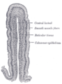 Vertical section of a villus from the dog's small intestine. X 80. (Simple columnar epithelium labeled at right, third from top.)
 -
 Transverse section of a villus, from the humanMuscle cellscut across.
 -
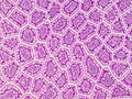 Cross-section histology of small intestinal villi of the humanterminal ileum.
 -
 MicroCT-based volume projection of the jejunal mucosa of a chicken. Virtual volume block with vertically truncated villi in oblique view. Scalebar = 0.2 mm.
 -
 MicroCT-based volume projection of the jejunal mucosa of a chicken. Virtual horizontal cut through villi. Scalebar = 0.2 mm.


Function
There, the villi and the microvilli increase intestinal absorptive surface area approximately 40-fold and 600-fold, respectively, providing exceptionally efficient absorption of nutrients in the lumen.[2]
There are also
Villi are specialized for absorption in the small intestine as they have a thin wall, one cell thick, which enables a shorter diffusion path. They have a large surface area so there will be more efficient absorption of fatty acids and glycerol into the blood stream. They have a rich blood supply to keep a concentration gradient.[3]
-
 Structure of a villus (see reference quoted in text)

Clinical significance
Villous atrophy

In diseases of the small intestine the villi can become flattened due to the effects of inflammation, and the villi can sometimes disappear. This deterioration is known as villous atrophy, and is often a feature of coeliac disease.[4]
Additional images
-
 Microvilli(shaggy hair) show electron dense plaques (open arrow) at their apices.

References
- ^ "Paneth cells (Cytokines & Cells Encyclopedia - COPE)".
- ISBN 978-1-4419-0199-6.
- ^ "Digestion: Digestive System, Enzymes, Absorption in the Small Intestine". Archived from the original on 2016-11-18. Retrieved 2014-12-30.
- ^ "Causes". Coeliac UK. Retrieved 12 July 2020.
Further reading
- C. W. Chan, Y. K. Leung and K. W. Chan (2014). "Microscopic anatomy of the vasculature of the human intestinal villus - a study with review". European Journal of Anatomy, 18 (4): 291–301.
| National | |
|---|---|
| Other | |