
Kidney
| Kidneys | |
|---|---|
 The kidneys lie in the retroperitoneal space behind the abdomen, and act to filter blood to create urine | |
 View of the kidneys from behind, showing their blood supply and drainage | |
| Details | |
| System | Urinary system and endocrine system |
| Artery | Renal artery |
| Vein | Renal vein |
| Nerve | Renal plexus |
| Identifiers | |
| Latin | ren |
| Greek | nephros |
| MeSH | D007668 |
| TA98 | A08.1.01.001 |
| TA2 | 3358 |
| FMA | 7203 |
| Anatomical terminology | |
In humans, the kidneys are two reddish-brown bean-shaped blood-filtering
The kidney participates in the control of the volume of various
The word “renal” is an adjective meaning “relating to the kidneys”, and its roots are French or late Latin. Whereas according to some opinions, "renal" should be replaced with "kidney" in scientific writings such as "kidney artery", other experts have advocated preserving the use of "renal" as appropriate including in "renal artery".[8]
Structure
In humans, the kidneys are located high in the
The human kidney is a bean-shaped structure with a
The superior pole of the right kidney is adjacent to the liver. For the left kidney, it is next to the spleen. Both, therefore, move down upon inhalation.
| Sex | Weight, standard reference range | |
| Right kidney | Left kidney | |
| Male[15] | 80–160 g (2+3⁄4–5+3⁄4 oz) | 80–175 g (2+3⁄4–6+1⁄4 oz) |
| Female[16] | 40–175 g (1+1⁄2–6+1⁄4 oz) | 35–190 g (1+1⁄4–6+3⁄4 oz) |
A Danish study measured the median renal length to be 11.2 cm (4+7⁄16 in) on the left side and 10.9 cm (4+5⁄16 in) on the right side in adults. Median renal volumes were 146 cm3 (8+15⁄16 cu in) on the left and 134 cm3 (8+3⁄16 cu in) on the right.[17]
Gross anatomy
The functional substance, or
The tip, or
The kidneys possess no overtly moving structures.
-
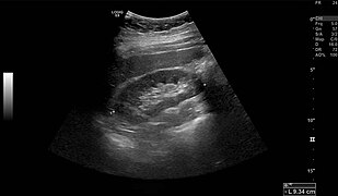 Normal adult right kidney as seen onabdominal ultrasoundwith a pole to pole measurement of 9.34 cm
 -

-
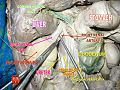
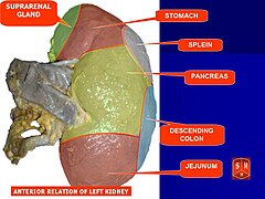 Image showing the structures that the kidney lies near
 -
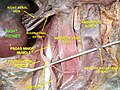
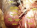
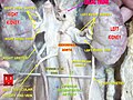
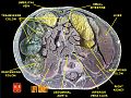
 Cross-section through a cadaveric specimen showing the position of the kidneys


Blood supply
The kidneys receive blood from the
Blood drains from the kidneys, ultimately into the
Nerve supply
The kidney and nervous system communicate via the renal plexus, whose fibers course along the renal arteries to reach each kidney.[22] Input from the sympathetic nervous system triggers vasoconstriction in the kidney, thereby reducing renal blood flow.[22] The kidney also receives input from the parasympathetic nervous system, by way of the renal branches of the vagus nerve; the function of this is yet unclear.[22][23] Sensory input from the kidney travels to the T10–11 levels of the spinal cord and is sensed in the corresponding dermatome.[22] Thus, pain in the flank region may be referred from corresponding kidney.[22]
Microanatomy
Renal
- Kidney glomerulus parietal cell
- Kidney glomerulus podocyte
- Kidney proximal tubule brush border cell
- Loop of Henle thin segment cell
- Thick ascending limbcell
- Kidney distal tubule cell
- Collecting duct principal cell
- Collecting duct intercalated cell
- Interstitial kidney cells
Gene and protein expression
In humans, about 20,000 protein coding genes are expressed in human cells and almost 70% of these genes are expressed in normal, adult kidneys.
Development
The mammalian kidney develops from intermediate mesoderm. Kidney development, also called nephrogenesis, proceeds through a series of three successive developmental phases: the pronephros, mesonephros, and metanephros. The metanephros are primordia of the permanent kidney.[28]
Function

The kidneys excrete a variety of waste products produced by metabolism into the urine. The microscopic structural and functional unit of the kidney is the nephron. It processes the blood supplied to it via filtration, reabsorption, secretion and excretion; the consequence of those processes is the production of urine. These include the nitrogenous wastes urea, from protein catabolism, and uric acid, from nucleic acid metabolism. The ability of mammals and some birds to concentrate wastes into a volume of urine much smaller than the volume of blood from which the wastes were extracted is dependent on an elaborate countercurrent multiplication mechanism. This requires several independent nephron characteristics to operate: a tight hairpin configuration of the tubules, water and ion permeability in the descending limb of the loop, water impermeability in the ascending loop, and active ion transport out of most of the ascending limb. In addition, passive countercurrent exchange by the vessels carrying the blood supply to the nephron is essential for enabling this function.
The kidney participates in whole-body
Formation of urine

Filtration
Filtration, which takes place at the renal corpuscle, is the process by which cells and large proteins are retained while materials of smaller molecular weights are[29] filtered from the blood to make an ultrafiltrate that eventually becomes urine. The adult human kidney generates approximately 180 liters of filtrate a day, most of which is reabsorbed.[30] The normal range for a twenty four hour urine volume collection is 800 to 2,000 milliliters per day.[31] The process is also known as hydrostatic filtration due to the hydrostatic pressure exerted on the capillary walls.
Reabsorption

Reabsorption is the transport of molecules from this ultrafiltrate and into the peritubular capillary. It is accomplished via selective receptors on the luminal cell membrane. Water is 55% reabsorbed in the proximal tubule. Glucose at normal plasma levels is completely reabsorbed in the proximal tubule. The mechanism for this is the Na+/glucose cotransporter. A plasma level of 350 mg/dL will fully saturate the transporters and glucose will be lost in the urine. A plasma glucose level of approximately 160 is sufficient to allow glucosuria, which is an important clinical clue to diabetes mellitus.
Amino acids are reabsorbed by sodium dependent transporters in the proximal tubule. Hartnup disease is a deficiency of the tryptophan amino acid transporter, which results in pellagra.[32]
| Location of Reabsorption | Reabsorbed nutrient | Notes |
|---|---|---|
| Early proximal tubule | Glucose (100%), amino acids (100%), bicarbonate (90%), Na+ (65%), Cl− (65%), phosphate (65%) and H2O (65%) |
|
| Thin descending loop of Henle | H2O |
|
| Thick ascending loop of Henle | Na+ (10–20%), K+, Cl−; indirectly induces para cellular reabsorption of Mg2+, Ca2+ |
|
| Early distal convoluted tubule | Na+, Cl− |
|
| Collecting tubules | Na+(3–5%), H2O |
|
| Examples of substances that are reabsorbed in the kidneys, and the hormones that influence those processes.[32] | ||
Secretion
Secretion is the reverse of reabsorption: molecules are transported from the peritubular capillary through the interstitial fluid, then through the renal tubular cell and into the ultrafiltrate.
Excretion
The last step in the processing of the ultrafiltrate is excretion: the ultrafiltrate passes out of the nephron and travels through a tube called the collecting duct, which is part of the collecting duct system, and then to the ureters where it is renamed urine. In addition to transporting the ultrafiltrate, the collecting duct also takes part in reabsorption.
Hormone secretion
The kidneys secrete a variety of
Blood pressure regulation
Although the kidney cannot directly sense blood, long-term regulation of
Acid–base balance
The two organ systems that help regulate the body's acid–base balance are the kidneys and lungs. Acid–base homeostasis is the maintenance of pH around a value of 7.4. The lungs are the part of respiratory system which helps to maintain acid–base homeostasis by regulating carbon dioxide (CO2) concentration in the blood. The respiratory system is the first line of defense when the body experiences and acid–base problem. It attempts to return the body pH to a value of 7.4 by controlling the respiratory rate. When the body is experiencing acidic conditions, it will increase the respiratory rate which in turn drives off CO2 and decreases the H+ concentration, therefore increasing the pH. In basic conditions, the respiratory rate will slow down so that the body holds onto more CO2 and increases the H+ concentration and decreases the pH.[citation needed]
The kidneys have two cells that help to maintain acid-base homeostasis: intercalated A and B cells. The intercalated A cells are stimulated when the body is experiencing acidic conditions. Under acidic conditions, the high concentration of CO2 in the blood creates a gradient for CO2 to move into the cell and push the reaction HCO3 + H ↔ H2CO3 ↔ CO2 + H2O to the left. On the luminal side of the cell there is a H+ pump and a H/K exchanger. These pumps move H+ against their gradient and therefore require ATP. These cells will remove H+ from the blood and move it to the filtrate which helps to increase the pH of the blood. On the basal side of the cell there is a HCO3/Cl exchanger and a Cl/K co-transporter (facilitated diffusion). When the reaction is pushed to the left it also increases the HCO3 concentration in the cell and HCO3 is then able to move out into the blood which additionally raises the pH. The intercalated B cell responds very similarly, however, the membrane proteins are flipped from the intercalated A cells: the proton pumps are on the basal side and the HCO3/Cl exchanger and K/Cl co-transporter are on the luminal side. They function the same, but now release protons into the blood to decrease the pH.[citation needed]
Regulation of osmolality
The kidneys help maintain the water and salt level of the body. Any significant rise in
Measuring function
Various calculations and methods are used to try to measure kidney function.
Clinical significance
There are many causes of
Medical terms related to the kidneys commonly use terms such as renal and the prefix nephro-. The
Acquired Disease
- Diabetic nephropathy
- Glomerulonephritis
- Hydronephrosis is the enlargement of one or both of the kidneys caused by obstruction of the flow of urine.
- Interstitial nephritis
- Kidney stones (nephrolithiasis) are a relatively common and particularly painful disorder. A chronic condition can result in scars to the kidneys. The removal of kidney stones involves ultrasoundtreatment to break up the stones into smaller pieces, which are then passed through the urinary tract. One common symptom of kidney stones is a sharp to disabling pain in the middle and sides of the lower back or groin.
- Kidney tumour
- Wilms tumor
- Renal cell carcinoma
- Lupus nephritis
- Minimal change disease
- In nephrotic syndrome, the glomerulus has been damaged so that a large amount of protein in the blood enters the urine. Other frequent features of the nephrotic syndrome include swelling, low serum albumin, and high cholesterol.
- Pyelonephritis is infection of the kidneys and is frequently caused by complication of a urinary tract infection.
- Kidney failure
- Acute kidney failure
- Stage 5 Chronic Kidney Disease
- Renal artery stenosis
- Renovascular hypertension
Kidney injury and failure
Generally, humans can live normally with just one kidney, as one has more functioning renal tissue than is needed to survive. Only when the amount of functioning kidney tissue is greatly diminished does one develop chronic kidney disease. Renal replacement therapy, in the form of dialysis or kidney transplantation, is indicated when the glomerular filtration rate has fallen very low or if the renal dysfunction leads to severe symptoms.[36]
Dialysis

Dialysis is a treatment that substitutes for the function of normal kidneys. Dialysis may be instituted when approximately 85%–90% of kidney function is lost, as indicated by a glomerular filtration rate (GFR) of less than 15. Dialysis removes metabolic waste products as well as excess water and sodium (thereby contributing to regulating blood pressure); and maintains many chemical levels within the body. Life expectancy is 5–10 years for those on dialysis; some live up to 30 years. Dialysis can occur via the blood (through a catheter or arteriovenous fistula), or through the peritoneum (peritoneal dialysis) Dialysis is typically administered three times a week for several hours at free-standing dialysis centers, allowing recipients to lead an otherwise essentially normal life.[37]
Congenital disease
- Congenital hydronephrosis
- Congenital obstruction of urinary tract
- Duplex kidneys, or double kidneys, occur in approximately 1% of the population. This occurrence normally causes no complications, but can occasionally cause urinary tract infections.[38][39]
- Duplicated ureter occurs in approximately one in 100 live births
- Horseshoe kidney occurs in approximately one in 400 live births
- Nephroblastoma(Syndromic Wilm's tumour)
- Nutcracker syndrome
- Polycystic kidney disease
- Autosomal dominant polycystic kidney disease affects patients later in life. Approximately one in 1000 people will develop this condition
- Autosomal recessive polycystic kidney disease is far less common, but more severe, than the dominant condition. It is apparent in utero or at birth.
- Renal agenesis. Failure of one kidney to form occurs in approximately one in 750 live births. Failure of both kidneys to form used to be fatal; however, medical advances such as amnioinfusion therapy during pregnancy and peritoneal dialysis have made it possible to stay alive until a transplant can occur.
- Renal dysplasia
- Unilateral small kidney
- Multicystic dysplastic kidney occurs in approximately one in every 2400 live births
- Ureteropelvic Junction Obstruction or UPJO; although most cases are congenital, some are acquired.[40]
Diagnosis
Many renal diseases are diagnosed on the basis of a detailed medical history, and physical examination.[41] The medical history takes into account present and past symptoms, especially those of kidney disease; recent infections; exposure to substances toxic to the kidney; and family history of kidney disease.
Imaging
Renal ultrasonography is essential in the diagnosis and management of kidney-related diseases.[44] Other modalities, such as CT and MRI, should always be considered as supplementary imaging modalities in the assessment of renal disease.[44]
Biopsy
The role of the renal biopsy is to diagnose renal disease in which the etiology is not clear based upon noninvasive means (clinical history, past medical history, medication history, physical exam, laboratory studies, imaging studies). In general, a renal pathologist will perform a detailed morphological evaluation and integrate the morphologic findings with the clinical history and laboratory data, ultimately arriving at a pathological diagnosis. A renal
Ideally, multiple core sections are obtained and evaluated for adequacy (presence of glomeruli) intraoperatively. A pathologist/pathology assistant divides the specimen(s) for submission for light microscopy, immunofluorescence microscopy and electron microscopy.
The pathologist will examine the specimen using light microscopy with multiple staining techniques (hematoxylin and eosin/H&E, PAS, trichrome, silver stain) on multiple level sections. Multiple immunofluorescence stains are performed to evaluate for antibody, protein and complement deposition. Finally, ultra-structural examination is performed with electron microscopy and may reveal the presence of electron-dense deposits or other characteristic abnormalities that may suggest an etiology for the patient's renal disease.
Other animals
In the majority of vertebrates, the
In the most primitive vertebrates, the
The kidneys of
Birds have relatively large, elongated kidneys, each of which is divided into three or more distinct lobes. The lobes consists of several small, irregularly arranged, lobules, each centred on a branch of the ureter. Birds have small glomeruli, but about twice as many nephrons as similarly sized mammals.[45]
The human kidney is fairly typical of that of mammals. Distinctive features of the mammalian kidney, in comparison with that of other vertebrates, include the presence of the renal pelvis and renal pyramids and a clearly distinguishable cortex and medulla. The latter feature is due to the presence of elongated loops of Henle; these are much shorter in birds, and not truly present in other vertebrates (although the nephron often has a short intermediate segment between the convoluted tubules). It is only in mammals that the kidney takes on its classical "kidney" shape, although there are some exceptions, such as the multilobed reniculate kidneys of pinnipeds and cetaceans.[45]
Evolutionary adaptation
Kidneys of various animals show evidence of evolutionary adaptation and have long been studied in ecophysiology and comparative physiology. Kidney morphology, often indexed as the relative medullary thickness, is associated with habitat aridity among species of mammals[47] and diet (e.g., carnivores have only long loops of Henle).[34]
Society and culture
Significance
Egyptian
In ancient Egypt, the kidneys, like the heart, were left inside the mummified bodies, unlike other organs which were removed. Comparing this to the biblical statements, and to drawings of human body with the heart and two kidneys portraying a set of scales for weighing justice, it seems that the Egyptian beliefs had also connected the kidneys with judgement and perhaps with moral decisions.[48]
Hebrew
According to studies in modern and ancient Hebrew, various body organs in humans and animals served also an emotional or logical role, today mostly attributed to the
(Berakhoth 61.a) states that one of the two kidneys counsels what is good, and the other evil.In the sacrifices offered at the biblical Tabernacle and later on at the temple in Jerusalem, the priests were instructed[50] to remove the kidneys and the adrenal gland covering the kidneys of the sheep, goat and cattle offerings, and to burn them on the altar, as the holy part of the "offering for God" never to be eaten.[51]
India: Ayurvedic system
In ancient India, according to the Ayurvedic medical systems, the kidneys were considered the beginning of the excursion channels system, the 'head' of the Mutra Srotas, receiving from all other systems, and therefore important in determining a person's health balance and temperament by the balance and mixture of the three 'Dosha's – the three health elements: Vatha (or Vata) – air, Pitta – bile, and Kapha – mucus. The temperament and health of a person can then be seen in the resulting color of the urine.[52]
Modern Ayurveda practitioners, a practice which is characterized as pseudoscience,[53] have attempted to revive these methods in medical procedures as part of Ayurveda Urine therapy.[54] These procedures have been called "nonsensical" by skeptics.[55]
Medieval Christianity
The Latin term renes is related to the English word "reins", a synonym for the kidneys in
History
Kidney stones have been identified and recorded about as long as written historical records exist.[59] The urinary tract including the ureters, as well as their function to drain urine from the kidneys, has been described by Galen in the second century AD.[60]
The first to examine the ureter through an internal approach, called ureteroscopy, rather than surgery was
Additional images
-
 Right kidney
 -
 Kidney
 -
 Right kidney
 -
 Right kidney
 -
 Left kidney
 -
 Kidneys
 -
 Left kidney


See also
- Artificial kidney
- Holonephros
- Nephromegaly
- Organ donation
- Organ harvesting
- Pelvic kidney
- World Kidney Day
- List of distinct cell types in the adult human body
References
Citations
- ^ "Kidneys: Anatomy, Function, Health & Conditions". Cleveland Clinic. Archived from the original on 2023-06-29. Retrieved 2023-07-13.
- ISBN 978-1-316-61398-6. Archivedfrom the original on 2023-04-04. Retrieved 2023-08-16.
- ISBN 978-0-12-415765-1.
- ^ Lote CJ (2012). Principles of Renal Physiology, 5th edition. Springer. p. 21.
- ^ Mescher AL (2016). Junqueira's Basic Histology, 14th edition. Lange. p. 393.
- S2CID 199519437.
- ISBN 978-0-7216-0187-8.
- PMID 33713333.
- ^ "HowStuffWorks How Your Kidney Works". 2001-01-10. Archived from the original on 2012-11-05. Retrieved 2012-08-09.
- ^ "Kidneys Location Stock Illustration". Archived from the original on 2013-09-27.
- ^ "Kidney". BioPortfolio Ltd. Archived from the original on 10 February 2008.
- PMID 20030823.
- ^ Dragomir A, Hjortberg M, Romans GM. Bålens ytanatomy [Superficial anatomy of the trunk]. Section for human anatomy at the Department of Medical Biology, Uppsala University, Sweden (Report) (in Swedish).
- ^ "Renal system". Britannica. Archived from the original on 2022-05-31. Retrieved 2022-05-22.
- S2CID 32174574.
- S2CID 25319215.
- PMID 8416654.
- ^ ISBN 978-1-4160-2328-9.
- ISBN 978-0-521-87702-2.
- ISBN 978-1-107-11047-2, archivedfrom the original on 2018-06-17, retrieved 2022-06-25
- ISBN 9780134320762.
- ^ ISBN 978-0-12-722441-1. Archivedfrom the original on 2023-08-17. Retrieved 2020-10-19.
- PMID 5056657.
- Wikidata Q27013996.
- ^ "The human proteome in kidney – The Human Protein Atlas". www.proteinatlas.org. Archived from the original on 2017-09-22. Retrieved 2017-09-22.
- S2CID 802377.
- PMID 25551756.
- ISBN 978-0-323-03649-8.
- ISBN 978-0-323-38930-3.
- ISBN 978-0-12-381463-0. Archivedfrom the original on 2023-07-22. Retrieved 2022-07-28.
- ^ "Urine 24-hour volume". mountsinai. Archived from the original on 21 November 2022. Retrieved 21 November 2022.
- ^ a b Le, Tao. First Aid for the USMLE Step 1 2013. New York: McGraw-Hill Medical, 2013. Print.
- PMID 8010757.
- ^ .
- ISBN 978-0-13-981176-0.
- (PDF) from the original on 2022-05-17. Retrieved 2022-05-22.
- ^ "Dialysis". National Kidney Foundation. 2015-12-24. Archived from the original on 2017-09-26. Retrieved 8 November 2017.
- ^ Sample I (2008-02-19). "How many people have four kidneys?". The Guardian. London. Archived from the original on 2016-08-17. Retrieved 2016-12-19.
- ^ "Kidneys Fail, Girl Survives with Spare Parts". Abcnews.go.com. 2010-05-18. Archived from the original on 2010-05-21. Retrieved 2011-01-03.
- )
- ^ Gaitonde DY (15 December 2017). "Chronic Kidney Disease: Detection and Evaluation". Am Fam Physician. 12 (96): 776–783. Archived from the original on 26 February 2021. Retrieved 1 March 2021.
- ^ a b Post TW, Rose BD (December 2012). Curhan GC, Sheridan AM (eds.). "Diagnostic Approach to the Patient With Acute Kidney Injury (Acute Kidney Failure) or Chronic Kidney Disease". www.uptodate.com. Archived from the original on 2015-11-10. Retrieved 2016-12-19.
- ^ "KDIGO 2012 Clinical Practice Guideline for the Evaluation and Management of Chronic Kidney Disease". Kidney Int Suppl. 3: 1–150. 2013. Archived from the original on 2019-05-01. Retrieved 2021-01-25.
- ^
- ^ ISBN 978-0-03-910284-5.
- PMID 24823278.
- S2CID 12420368. Archived from the original(PDF) on 2010-06-17. Retrieved 2009-03-28.
- S2CID 35305403.
- ^ "Body Part Metaphors in Biblical Hebrew by David Steinberg". March 22, 2003. Archived from the original on March 22, 2003. Retrieved July 21, 2019.
- ^ Leviticus 3: 4, 10 and 15
- ^ ie Deut 3:4,9,10,15... or the Babylonian Talmud, Bechorot (39a) Ch6:Tr2...
- ^ "What is Vata Dosha? Tips and diet for balancing vata | CA College of Ayurveda". www.ayurvedacollege.com. 7 April 2010. Archived from the original on 9 November 2019. Retrieved July 21, 2019.
- ^ List of topics characterized as pseudoscience, according to the American Medical Association's Report 12 of the Council of Scientific Affairs (A-97) and claims by skeptics Archived 2016-08-10 at the Wayback Machine ('The Skeptics Dictionary' website)
- PMID 22131762.
- ^ Barrett S. "A Few Thoughts on Ayurvedic Mumbo-Jumbo". Archived from the original on 2020-09-29. Retrieved 2022-05-22. M.D, head of the National Council Against Health Fraud NGO and owner of the QuackWatch website.
- ISBN 978-0-300-09396-4.
- ISBN 978-3-8055-6499-1. International Association for the History of Nephrology Congress, Reprint of American Journal of Nephrology; v. 14, no. 4–6, 1994.
- ^ intertextual.bible/text/revelation-2.23-berakhot-119.29, archived from the original on 2022-12-15, retrieved 2022-12-15
- ^ PMID 24348156.
- PMID 21805756.
General and cited references
- Barrett KE, Barman SM, Yuan JX, Brooks H (2019). Ganong's review of medical physiology (26th ed.). New York. OCLC 1076268769.)
{{cite book}}: CS1 maint: location missing publisher (link