Lamina affixa
| Lamina affixa | |
|---|---|
Middle cerebellar peduncles | |
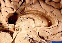
 Human brain left dissected midsagittal view (Lamina affixa is #10) | |
| Details | |
| Identifiers | |
| Latin | lamina affixa |
| TA98 | A14.1.09.276 |
| TA2 | 5650 |
| FMA | 83709 |
| Anatomical terms of neuroanatomy] | |
Lamina affixa is a layer of epithelium growing on the surface of the
superior choroid vein. The torn edge of this plexus is called the tela choroidea.[1]
On the surface of the terminal vein is a narrow white band, named the lamina affixa.
GDF-15/MIC-1 has been observed in lamina affixa cells.[2]
References
![]() This article incorporates text in the public domain from page 838 of the 20th edition of Gray's Anatomy (1918)
This article incorporates text in the public domain from page 838 of the 20th edition of Gray's Anatomy (1918)
- ISBN 978-1-4160-6257-8.
- S2CID 40302340.
External links
