
Larynx
| Larynx | |
|---|---|
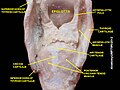
 Anatomy of the larynx, anterolateral view | |
| Details | |
| Pronunciation | /ˈlærɪŋks/ |
| Identifiers | |
| Latin | larynx |
| MeSH | D007830 |
| TA98 | A06.2.01.001 |
| TA2 | 3184 |
| FMA | 55097 |
| Anatomical terminology | |
The larynx (
Structure

The triangle-shaped larynx consists largely of cartilages that are attached to one another, and to surrounding structures, by muscles or by fibrous and elastic tissue components. The larynx is lined by a
Location
In adult humans, the larynx is found in the

Cartilages


There are nine cartilages, three unpaired and three paired (3 pairs=6), that support the mammalian larynx and form its skeleton.
Unpaired cartilages:
- Thyroid cartilage: This forms the Adam's apple (also called the laryngeal prominence). It is usually larger in males than in females. The thyrohyoid membrane is a ligament associated with the thyroid cartilage that connects it with the hyoid bone. It supports the front portion of the larynx.
- median cricothyroid ligamentconnects the cricoid cartilage to the thyroid cartilage.
- Epiglottis: A large, spoon-shaped piece of elastic cartilage. During swallowing, the pharynx and larynx rise. Elevation of the pharynx widens it to receive food and drink; elevation of the larynx causes the epiglottis to move down and form a lid over the glottis, closing it off.
Paired cartilages:
- Arytenoid cartilages: Of the paired cartilages, the arytenoid cartilages are the most important because they influence the position and tension of the vocal cords. These are triangular pieces of mostly hyaline cartilage located at the posterosuperior border of the cricoid cartilage.
- Corniculate cartilages: Horn-shaped pieces of elastic cartilage located at the apex of each arytenoid cartilage.
- Cuneiform cartilages: Club-shaped pieces of elastic cartilage located anterior to the corniculate cartilages.
Muscles
The muscles of the larynx are divided into intrinsic and extrinsic muscles. The extrinsic muscles act on the region and pass between the larynx and parts around it but have their origin elsewhere; the intrinsic muscles are confined entirely within the larynx and have their origin and insertion there.[4]
The intrinsic muscles are divided into respiratory and the phonatory muscles (the muscles of
Intrinsic
The intrinsic laryngeal muscles are responsible for controlling sound production.
- Cricothyroid muscle lengthen and tense the vocal cords.
- Posterior cricoarytenoid muscles abduct and externally rotate the arytenoid cartilages, resulting in abducted vocal cords.
- Lateral cricoarytenoid muscles adduct and internally rotate the arytenoid cartilages, increase medial compression.
- Transverse arytenoid muscle adduct the arytenoid cartilages, resulting in adducted vocal cords.[5]
- Oblique arytenoid muscles narrow the laryngeal inletby constricting the distance between the arytenoid cartilages.
- Thyroarytenoid muscles narrow the laryngeal inlet, shortening the vocal cords, and lowering voice pitch. The internal thyroarytenoid is the portion of the thyroarytenoid that vibrates to produce sound.
Notably the only muscle capable of separating the vocal cords for normal breathing is the posterior cricoarytenoid. If this muscle is incapacitated on both sides, the inability to pull the vocal cords apart (abduct) will cause difficulty breathing. Bilateral injury to the recurrent laryngeal nerve would cause this condition. It is also worth noting that all muscles are innervated by the recurrent laryngeal branch of the vagus except the cricothyroid muscle, which is innervated by the external laryngeal branch of the superior laryngeal nerve (a branch of the vagus).
Additionally, intrinsic laryngeal muscles present a constitutive
Extrinsic
The extrinsic laryngeal muscles support and position the larynx within the mid-cervical cereal region.

- Sternothyroid muscles depress the larynx. (Innervated by ansa cervicalis)
- Omohyoid muscles depress the larynx. (Ansa cervicalis)
- Sternohyoid muscles depress the larynx. (Ansa cervicalis)
- Inferior constrictor muscles. (CN X)
- Thyrohyoid muscles elevates the larynx. (C1)
- Digastricelevates the larynx. (CN V3, CN VII)
- Stylohyoidelevates the larynx. (CN VII)
- Mylohyoid elevates the larynx. (CN V3)
- Geniohyoidelevates the larynx. (C1)
- Hyoglossus elevates the larynx. (CN XII)
- Genioglossus elevates the larynx. (CN XII)
Nerve supply
The larynx is
Injury to the external branch of the superior laryngeal nerve causes weakened phonation because the vocal cords cannot be tightened. Injury to one of the recurrent laryngeal nerves produces
Development
In newborn infants, the larynx is initially at the level of the C2–C3 vertebrae, and is further forward and higher relative to its position in the adult body.[8] The larynx descends as the child grows.[9][10]
Laryngeal cavity
| Laryngeal cavity | |
|---|---|
 Sagittal section of the larynx and upper part of the trachea. | |
 Coronal section of larynx and upper part of trachea. | |
| Details | |
| Identifiers | |
| Latin | cavitas laryngis |
| MeSH | D007830 |
| TA98 | A06.2.01.001 |
| TA2 | 3184 |
| FMA | 55097 |
| Anatomical terminology | |
The laryngeal cavity (cavity of the larynx) extends from the laryngeal inlet downwards to the lower border of the cricoid cartilage where it is continuous with that of the trachea.[11][12]
It is divided into two parts by the projection of the
The portion of the cavity of the larynx above the vocal folds is called the laryngeal vestibule; it is wide and triangular in shape, its base or anterior wall presenting, however, about its center the backward projection of the tubercle of the epiglottis.
It contains the vestibular folds, and between these and the vocal folds are the laryngeal ventricles.
The portion below the vocal folds is called the infraglottic cavity. It is at first of an elliptical form, but lower down it widens out, assumes a circular form, and is continuous with the tube of the trachea.
Function
Sound generation
Sound is generated in the larynx, and that is where pitch and volume are manipulated. The strength of expiration from the lungs also contributes to loudness.
Manipulation of the larynx is used to generate a source sound with a particular fundamental frequency, or pitch. This source sound is altered as it travels through the vocal tract, configured differently based on the position of the tongue, lips, mouth, and pharynx. The process of altering a source sound as it passes through the filter of the vocal tract creates the many different vowel and consonant sounds of the world's languages as well as tone, certain realizations of stress and other types of linguistic prosody. The larynx also has a similar function to the lungs in creating pressure differences required for sound production; a constricted larynx can be raised or lowered affecting the volume of the oral cavity as necessary in glottalic consonants.
The vocal cords can be held close together (by adducting the arytenoid cartilages) so that they vibrate (see phonation). The muscles attached to the arytenoid cartilages control the degree of opening. Vocal cord length and tension can be controlled by rocking the thyroid cartilage forward and backward on the cricoid cartilage (either directly by contracting the cricothyroids or indirectly by changing the vertical position of the larynx), by manipulating the tension of the muscles within the vocal cords, and by moving the arytenoids forward or backward. This causes the pitch produced during phonation to rise or fall. In most males the vocal cords are longer and have a greater mass than most females' vocal cords, producing a lower pitch.
The vocal apparatus consists of two pairs of folds, the vestibular folds (false vocal cords) and the true vocal cords. The vestibular folds are covered by respiratory epithelium, while the vocal cords are covered by stratified squamous epithelium. The vestibular folds are not responsible for sound production, but rather for resonance. The exceptions to this are found in Tibetan chanting and Kargyraa, a style of Tuvan throat singing. Both make use of the vestibular folds to create an undertone. These false vocal cords do not contain muscle, while the true vocal cords do have skeletal muscle.
Other

The most important role of the larynx is its protective function, the prevention of foreign objects from entering the lungs by
Another important role of the larynx is abdominal fixation, a kind of Valsalva maneuver in which the lungs are filled with air in order to stiffen the thorax so that forces applied for lifting can be translated down to the legs. This is achieved by a deep inhalation followed by the adduction of the vocal cords. Grunting while lifting heavy objects is the result of some air escaping through the adducted vocal cords ready for phonation.[13]
Abduction of the vocal cords is important during physical exertion. The vocal cords are separated by about 8 mm (0.31 in) during normal respiration, but this width is doubled during forced respiration.[13]
During swallowing, elevation of the posterior portion of the tongue levers (inverts) the epiglottis over the glottis' opening to prevent swallowed material from entering the larynx which leads to the lungs, and provides a path for a food or liquid bolus to "slide" into the esophagus; the hyo-laryngeal complex is also pulled upwards to assist this process. Stimulation of the larynx by aspirated food or liquid produces a strong cough reflex to protect the lungs.
In addition, intrinsic laryngeal muscles are spared from some muscle wasting disorders, such as Duchenne muscular dystrophy, may facilitate the development of novel strategies for the prevention and treatment of muscle wasting in a variety of clinical scenarios. ILM have a calcium regulation system profile suggestive of a better ability to handle calcium changes in comparison to other muscles, and this may provide a mechanistic insight for their unique pathophysiological properties[6]
Clinical significance
Disorders
There are several things that can cause a larynx to not function properly.[14] Some symptoms are hoarseness, loss of voice, pain in the throat or ears, and breathing difficulties.
- Acute laryngitisis the sudden inflammation and swelling of the larynx. It is caused by the common cold or by excessive shouting. It is not serious.
- Chronic laryngitisis caused by smoking, dust, frequent yelling, or prolonged exposure to polluted air. It is much more serious than acute laryngitis.
- Presbylarynx is a condition in which age-related atrophy of the soft tissues of the larynx results in weak voice and restricted vocal range and stamina. Bowing of the anterior portion of the vocal colds is found on laryngoscopy.
- endotracheal tube.
- Polyps and vocal cord nodules are small bumps caused by prolonged exposure to tobacco smoke and vocal misuse, respectively.
- Two related types of squamous cell carcinoma and verrucous carcinoma, are strongly associated with repeated exposure to cigarette smoke and alcohol.
- Vocal cord paresis is weakness of one or both vocal cords that can greatly impact daily life.
- Idiopathic laryngeal spasm.
- Laryngopharyngeal reflux is a condition in which acid from the stomach irritates and burns the larynx. Similar damage can occur with gastroesophageal reflux disease (GERD).[15][16]
- Laryngomalacia is a very common condition of infancy, in which the soft, immature cartilage of the upper larynx collapses inward during inhalation, causing airway obstruction.
- Laryngeal perichondritis, the inflammation of the perichondriumof laryngeal cartilages, causing airway obstruction.
- Laryngeal paralysis is a condition seen in some mammals (including dogs) in which the larynx no longer opens as wide as required for the passage of air, and impedes respiration. In mild cases it can lead to exaggerated or "raspy" breathing or panting, and in serious cases can pose a considerable need for treatment.
- myonecrosis. The results further support the concept that abnormal calcium buffering is involved in these neuromuscular diseases.[17]
Treatments
Patients who have lost the use of their larynx are typically prescribed the use of an
Other animals
Pioneering work on the structure and evolution of the larynx was carried out in the 1920s by the British comparative anatomist
In contrast, though other species have low larynges, their tongues remain anchored in their mouths and their vocal tracts cannot produce the range of speech sounds of humans. The ability to lower the larynx transiently in some species extends the length of their vocal tract, which as Fitch showed creates the acoustic illusion that they are larger. Research at Haskins Laboratories in the 1960s showed that speech allows humans to achieve a vocal communication rate that exceeds the fusion frequency of the auditory system by fusing sounds together into syllables and words. The additional speech sounds that the human tongue enables us to produce, particularly [i], allow humans to unconsciously infer the length of the vocal tract of the person who is talking, a critical element in recovering the phonemes that make up a word.[24]
Non-mammals
Most
An example of a frog that possesses a larynx is túngara frog. While larynx is the main sound producing organ in túngara frogs, it serves a higher significance due to its contribution to mating call, which consist of two components: 'whine' and 'chuck'.[26] While 'whine' induces female phonotaxis and allows species recognition, 'chuck' increases mating attractiveness.[27] In particular, túngara frog produces 'chuck' by vibrating the fibrous mass attached to the larynx.[27]
Vocal folds are found only in mammals, and a few
History
The ancient Greek physician Galen first described the larynx, describing it as the "first and supremely most important instrument of the voice".[28]
Additional images
-
 Larynx. Deep dissection. Anterior view.
 -
 Larynx. Deep dissection. Posterior view.


See also
References
Notes
- PMID 30855790. Retrieved 2021-04-02.
The larynx is about 4 to 5cm in length and width, with a slightly shorter anterior-posterior diameter. It is smaller in women than men, and larger in adults than children owing to its growth in puberty. A larger larynx correlates with a deeper voice.
- ^ "Larynx Etymology". Online Etymology Dictionary. Retrieved 25 October 2015.
- ^ Knipe H. "Laryngeal cartilages". Radiology Reference Article. Radiopaedia.org.
- ISBN 9780071222075.
- ^ Collectively, the transverse and oblique arytenoids are known as the interarytenoids.
- ^ PMID 26109185.
- ^ S2CID 41968787.
- ^ "GERD and aspiration in the child: diagnosis and treatment". Grand Rounds Presentation. UTMB Dept. of Otolaryngology. February 23, 2005. Archived from the original on June 1, 2010. Retrieved June 16, 2010.
- ^ Laitman & Reidenberg 2009
- ^ Laitman, Noden & Van De Water 2006
- ^ "Pharynx" Emory University Anatomy Manual. Retrieved 2015-09-10.
- ^ "Chapter 53: The pharynx and larynx" Archived 2018-08-13 at the Wayback Machine Basic Human Anatomy. Retrieved 2015-09-10.
- ^ a b c Seikel, King & Drumright 2010, Nonspeech laryngeal function, pp. 223–225
- ^ Laitman & Reidenberg 1993
- ^ Laitman & Reidenberg 1997
- ^ Lipan, Reidenberg & Laitman 2006
- S2CID 25759998.
- ^ Helms D (December 2004). "Whispers on the Web - December 2004". Archived from the original on 2017-12-12. Retrieved 2019-08-06.
- ^ Communication after laryngectomy. YouTube. South East Coast Laryngectomy Support Groups (UK). 2011-03-09. Archived from the original on 2021-11-07. Retrieved 2013-03-14.
- ^ a b Only Human (2018-06-20). Speaking with a Dead Man's Voice by Organ Transplant Surgery | Only Human. Cineflix. YouTube. Retrieved 2019-08-06.
- S2CID 24360597.
- ^ Jensen B (January 21, 2011). "Rare transplant gives California woman a voice for the first time in a decade". Archived from the original on June 28, 2017. Retrieved January 13, 2015.
- ^ Johnson A (January 21, 2011). "Woman Finds Her Voice After Rare Transplant". Wall Street Journal. Retrieved 4 September 2012.
- ^ a b Lieberman 2006
- ^ a b Romer & Parsons 1977, pp. 214–215, 336
- S2CID 14153228.
- ^ ISBN 9780080453378.
- ISBN 978-91-7409-123-6.)
{{cite book}}: CS1 maint: location missing publisher (link
Sources
- Laitman JT, Noden DM, Van De Water TR (2006). "Formation of the larynx: from homeobox genes to critical periods". In Rubin JS, Sataloff RT, Korovin GS (eds.). Diagnosis & Treatment Voice Disorders. San Diego: Plural. pp. 3–20. OCLC 63279542.
- Laitman JT, Reidenberg JS (1993). "Specializations of the human upper respiratory and upper digestive systems as seen through comparative and developmental anatomy". Dysphagia. 8 (4): 318–325. S2CID 23308320.
- Laitman JT, Reidenberg JS (November 1997). "The human aerodigestive tract and gastroesophageal reflux: an evolutionary perspective". The American Journal of Medicine. 103 (5A): 2S–8S. PMID 9422615.
- Laitman JT, Reidenberg JS (2009). "The evolution of the human larynx: Nature's great experiment". In Fried MP, Ferlito A (eds.). The Larynx (3rd ed.). San Diego: Plural. pp. 19–38. OCLC 183609898.
- Lieberman P (2006). Toward an Evolutionary Biology of Language. Harvard University Press. OCLC 62766735.
- Lipan MJ, Reidenberg JS, Laitman JT (November 2006). "Anatomy of reflux: a growing health problem affecting structures of the head and neck". The Anatomical Record Part B: The New Anatomist. 289 (6): 261–270. PMID 17109421.
- Romer AS, Parsons TS (1977). The Vertebrate Body. Philadelphia, PA: Holt-Saunders International. ISBN 0-03-910284-X.
- Seikel JA, King DW, Drumright DG (2010). Anatomy & Physiology for Speech, Language, and Hearing (4th ed.). Delmar, NY: Cengage Learning. ISBN 978-1-4283-1223-4.