
Lateral geniculate nucleus
| Lateral geniculate nucleus | |
|---|---|
Terminal vein | |
| Identifiers | |
| Latin | corpus geniculatum laterale |
| Acronym(s) | LGN |
| NeuroNames | 352 |
| NeuroLex ID | birnlex_1662 |
| TA98 | A14.1.08.302 |
| TA2 | 5666 |
| FMA | 62209 |
| Anatomical terms of neuroanatomy] | |
In
The LGN receives information directly from the ascending
Structure

Both the left and right
M, P, K cells

| Type | Size* | RGC Source | Type of Information | Location | Response | Number |
| M: Magnocellular cells |
Large | Parasol cells | perception of movement, depth, and small differences in brightness | Layers 1 and 2 | rapid and transient | ? |
| P: Parvocellular cells (or "parvicellular") |
Small | Midget cells | perception of color and form (fine details) | Layers 3, 4, 5 and 6 | slow and sustained | ? |
| K: Koniocellular cells (or "interlaminar") |
Very small cell bodies | Bistratified cells | Between each of the M and P layers |
*Size describes the cell body and dendritic tree, though also can describe the receptive field
The magnocellular, parvocellular, and koniocellular layers of the LGN correspond with the similarly named types of retinal ganglion cells. Retinal P ganglion cells send axons to a parvocellular layer, M ganglion cells send axons to a magnocellular layer, and K ganglion cells send axons to a koniocellular layer.[5]: 269
Koniocellular cells are functionally and neurochemically distinct from M and P cells and provide a third channel to the visual cortex. They project their axons between the layers of the lateral geniculate nucleus where M and P cells project. Their role in visual perception is presently unclear; however, the koniocellular system has been linked with the integration of somatosensory system-proprioceptive information with visual perception[citation needed], and it may also be involved in color perception.[6]
The parvo- and magnocellular fibers were previously thought to dominate the Ungerleider–Mishkin
The other major retino–cortical visual pathway is the
Ipsilateral and contralateral layers
Both the LGN in the right hemisphere and the LGN in the left hemisphere receive input from each eye. However, each LGN only receives information from one half of the visual field.
- the eye on the same side (the ipsilateral eye) sends information to layers 2, 3 and 5
- the eye on the opposite side (the contralateral eye) sends information to layers 1, 4 and 6.
This description applies to the LGN of many primates, but not all. The sequence of layers receiving information from the ipsilateral and contralateral (opposite side of the head) eyes is different in the tarsier.[9] Some neuroscientists suggested that "this apparent difference distinguishes tarsiers from all other primates, reinforcing the view that they arose in an early, independent line of primate evolution".[10]
Input
The LGN receives input from the retina and many other brain structures, especially visual cortex.
The principal neurons in the LGN receive strong inputs from the retina. However, the retina only accounts for a small percentage of LGN input. As much as 95% of input in the LGN comes from the visual cortex, superior colliculus, pretectum, thalamic reticular nuclei, and local LGN interneurons. Regions in the brainstem that are not involved in visual perception also project to the LGN, such as the mesencephalic reticular formation, dorsal raphe nucleus, periaqueuctal grey matter, and the locus coeruleus.
Output
Information leaving the LGN travels out on the optic radiations, which form part of the retrolenticular portion of the internal capsule.
The axons that leave the LGN go to V1 visual cortex. Both the magnocellular layers 1–2 and the parvocellular layers 3–6 send their axons to layer 4 in V1. Within layer 4 of V1, layer 4cβ receives parvocellular input, and layer 4cα receives magnocellular input. However, the koniocellular layers, intercalated between LGN layers 1–6 send their axons primarily to the cytochrome-oxidase rich blobs of layers 2 and 3 in V1.[13] Axons from layer 6 of visual cortex send information back to the LGN.
Studies involving blindsight have suggested that projections from the LGN travel not only to the primary visual cortex but also to higher cortical areas V2 and V3. Patients with blindsight are phenomenally blind in certain areas of the visual field corresponding to a contralateral lesion in the primary visual cortex; however, these patients are able to perform certain motor tasks accurately in their blind field, such as grasping. This suggests that neurons travel from the LGN to both the primary visual cortex and higher cortex regions.[14]
Function in visual perception
The output of the LGN serves several functions.
Computations are achieved to determine the position of every major element in object space relative to the principal plane. Through subsequent motion of the eyes, a larger stereoscopic mapping of the visual field is achieved.[15]
It has been shown that while the retina accomplishes spatial decorrelation through center surround inhibition, the LGN accomplishes temporal decorrelation.[16] This spatial–temporal decorrelation makes for much more efficient coding. However, there is almost certainly much more going on.
Like other areas of the
Axiomatically determined functional models of LGN cells have been determined by Lindeberg [19][20] in terms of Laplacian of Gaussian kernels over the spatial domain in combination with temporal derivatives of either non-causal or time-causal scale-space kernels over the temporal domain. It has been shown that this theory both leads to predictions about receptive fields with good qualitative agreement with the biological receptive field measurements performed by DeAngelis et al.[21][22] and guarantees good theoretical properties of the mathematical receptive field model, including covariance and invariance properties under natural image transformations.[23][24] Specifically according to this theory, non-lagged LGN cells correspond to first-order temporal derivatives, whereas lagged LGN cells correspond to second-order temporal derivatives.
Color processing
The LGN is integral in the early steps of color processing, where opponent channels are created that compare signals between the different Photoreceptor cell types. The output of P-cells comprises red-green opponent signals. The output of M-cells does not include much color opponency, rather a sum of the red-green signal that evokes luminance. The output of K-cells comprises mostly blue-yellow opponent signals.[25]
Rodents
In rodents, the lateral geniculate nucleus contains the dorsal lateral geniculate nucleus (dLGN), the ventral lateral geniculate nucleus (vLGN), and the region in between called the intergeniculate leaflet (IGL). These are distinct subcortical nuclei with differences in function.
dLGN
The dorsolateral geniculate nucleus is the main division of the lateral geniculate body. The majority of input to the dLGN comes from the retina. It is laminated and shows retinotopic organization.[26]
vLGN
The ventrolateral geniculate nucleus has been found to be relatively large in several species such as lizards, rodents, cows, cats, and primates.[27] An initial cytoarchitectural scheme, which has been confirmed in several studies, suggests that the vLGN is divided into two parts. The external and internal divisions are separated by a group of fine fibers and a zone of thinly dispersed neurons. Additionally, several studies have suggested further subdivisions of the vLGN in other species.[28] For example, studies indicate that the cytoarchitecture of the vLGN in the cat differs from rodents. Although five subdivisions of the vLGN in the cat have been identified by some,[29] the scheme that divides the vLGN into three regions (medial, intermediate, and lateral) has been more widely accepted.
IGL
The intergeniculate leaflet is a relatively small area found dorsal to the vLGN. Earlier studies had referred to the IGL as the internal dorsal division of the vLGN. Several studies have described homologous regions in several species, including humans.[30]
The vLGN and IGL appear to be closely related based on similarities in neurochemicals, inputs and outputs, and physiological properties.
The vLGN and IGL have been reported to share many neurochemicals that are found concentrated in the cells, including neuropeptide Y, GABA, encephalin, and nitric oxide synthase. The neurochemicals serotonin, acetylcholine, histamine, dopamine, and noradrenaline have been found in the fibers of these nuclei.
Both the vLGN and IGL receive input from the retina, locus coreuleus, and raphe. Other connections that have been found to be reciprocal include the superior colliculus, pretectum, and hypothalamus, as well as other thalamic nuclei.
Physiological and behavioral studies have shown spectral-sensitive and motion-sensitive responses that vary with species. The vLGN and IGL seem to play an important role in mediating phases of the circadian rhythms that are not involved with light, as well as phase shifts that are light-dependent.[28]
Additional images
-
 Thalamus
 -
 Dissection of brain-stem. Lateral view.
 -
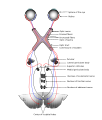 Scheme showing central connections of the optic nerves and optic tracts.
 -
 Thalamic nuclei
 -
 3D schematic representation of optic tracts
 -
 Brainstem. Posterior view.


References
- S2CID 6301290.
- ^ Goodale, M. & Milner, D. (2004)Sight unseen.Oxford University Press, Inc.: New York.
- ISBN 978-0-19-538115-3.
- ISBN 978-0205467242.
- ISBN 978-0878936953.
- PMID 19793973.
- ^ Goodale & Milner, 1993, 1995.
- ^ Nicholls J., et al. From Neuron to Brain: Fourth Edition. Sinauer Associates, Inc. 2001.
- PMID 8872317.
- PMID 16200648.
- ^ PMID 11804565.
- S2CID 29034684." and also "Kaas, J.H., and Huerta, M.F. 1988. The subcortical visual system of primates. In: Steklis H. D., Erwin J., editors. Comparative primate biology, vol 4: neurosciences. New York: Alan Liss, pp. 327–391.
- PMID 10845061.
- PMID 20574422.
- ^ Lindstrom, S. & Wrobel, A. (1990) Intracellular recordings from binocularly activated cells in the cats dorsal lateral geniculate nucleus Acta Neurobiol Exp vol 50, pp 61–70
- ^ Dawei W. Dong and Joseph J. Atick, Network–Temporal Decorrelation: A Theory of Lagged and Nonlagged Responses in the Lateral Geniculate Nucleus, 1995, pp. 159–178.
- PMID 16624964.
- PMID 22687612.
- PMID 24197240.
- PMID 33521348.
- S2CID 12827601.
- ^ G. C. DeAngelis and A. Anzai "A modern view of the classical receptive field: linear and non-linear spatio-temporal processing by V1 neurons. In: Chalupa, L.M., Werner, J.S. (eds.) The Visual Neurosciences, vol. 1, pp. 704–719. MIT Press, Cambridge, 2004.
- PMID 23894283.
- ^ T. Lindeberg "Covariance properties under natural image transformations for the generalized Gaussian derivative model for visual receptive fields", Frontiers in Computational Neuroscience, 17:1189949, 2023.
- ^ M. Ghodrati, S.-M. Khaligh-Razavi, S.R. Lehky, Towards building a more complex view of the lateral geniculate nucleus: recent advances in understanding its role, Prog. Neurobiol. 156:214–255, 2017.
- PMID 14687550.
- S2CID 28607983.
- ^ S2CID 20139828.
- S2CID 30586321.
- S2CID 33543381.
External links
- Malpeli J. Malpeli Lab Home Page. Retrieved September 1, 2004.
- Stained brain slice images which include the "lateral%20geniculate%20nucleus" at the BrainMaps project
- Atlas image: eye_38 at the University of Michigan Health System – "The Visual Pathway from Below"
- Stained brain slice images which include the "lgn" at the BrainMaps project
- MedicalMnemonics.com: 307 640
| Illusions |
| |
|---|---|---|
| Popular culture |
| |
| Related | ||