
Lateral pterygoid muscle
| Lateral pterygoid muscle | |
|---|---|
lateral pterygoid plate | |
| Insertion | Superior head: anterior side of the mandibular condyle. Inferior head: pterygoid fovea |
| Artery | pterygoid branches of maxillary artery |
| Nerve | lateral pterygoid nerve from mandibular nerve |
| Actions | depresses and protrudes mandible, side to side movement of mandible |
| Identifiers | |
| Latin | musculus pterygoideus lateralis, musculus pterygoideus externus |
| TA98 | A04.1.04.006 |
| TA2 | 2109 |
| FMA | 49015 |
| Anatomical terms of muscle] | |
The lateral pterygoid muscle (or external pterygoid muscle) is a muscle of
Structure
The lateral pterygoid muscle has an upper head and a lower head.[1][2]
- The upper head originates on the infratemporal surface and infratemporal crest of the greater wing of the fibrous capsule of the temporomandibular joint.
- The lower head originates on the lateral surface of the mandible.
It lies superior to the medial pterygoid muscle.
Blood supply
The lateral pterygoid muscle is supplied by pterygoid branches of the maxillary artery.[citation needed]
Nerve supply
The lateral pterygoid muscle is supplied by the lateral pterygoid nerve, a branch of the mandibular nerve (CN V3), itself a branch of the trigeminal nerve (CN V).
Function
The primary function of the lateral pterygoid muscle is to pull the head of the condyle out of the
Unlike the other three muscles of mastication, the lateral pterygoid alone can assist in depressing the mandible (opening the jaw). At the beginning of this action it is assisted by the
Clinical significance
The lateral pterygoid muscle may be involved in temporomandibular joint dysfunction.[1][2]
Additional images
-
 Sphenoid bone. Anterior and inferior surfaces.
 -
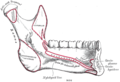 Mandible. Inner surface. Side view.
 -
 Plan of branches of internal maxillary artery.
 -
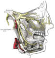 Distribution of the maxillary and mandibular nerves, and the submaxillary ganglion.


References
External links
- lesson4 at The Anatomy Lesson by Wesley Norman (Georgetown University) (musclesofmastication2)
- MedicalMnemonics.com: 70
- "Anatomy diagram: 25420.000-1". Roche Lexicon - illustrated navigator. Elsevier. Archived from the original on 2015-02-26.
- Cross section at tufts.edu