
Long thoracic nerve
| Long thoracic nerve | |
|---|---|
 Nerves of the left upper extremity. (Long thoracic labeled vertically at shoulder, to left of artery.) | |
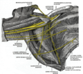
 The right brachial plexus with its short branches, viewed from in front. (Long thoracic labeled at center, third from top.) | |
| Details | |
| From | brachial plexus (C5-C7) |
| Innervates | serratus anterior muscle |
| Identifiers | |
| Latin | nervus thoracicus longus |
| TA98 | A14.2.03.012 |
| TA2 | 6410 |
| FMA | 65275 |
| Anatomical terms of neuroanatomy | |
The long thoracic nerve (also: external respiratory nerve of Bell or posterior thoracic nerve) is a branch of the
Structure
Origin
The long thoracic nerve arises from the
Course and relations
The long thoracic nerve descends through the cervicoaxillary canal. It is posterior to the brachial plexus,[3] and the axillary artery and vein.[4] This takes it deep to the clavicle.[2] It rests on the outer surface of the serratus anterior muscle. It extends along the side of the thorax to the lower border of the serratus anterior muscle, supplying fibres to each of the muscle's digitations.[5][6]
Function
The long thoracic nerve innervates the serratus anterior muscle.[1] It supplies filaments to each of its digitations (finger-like projections).[5][6]
Clinical significance
Due to its long, relatively superficial course, the long thoracic nerve is susceptible to injury, either through direct trauma or stretch.[7] Mechanisms of injury include:
- nerve lesions.[3]
- various sports injuries, typically occurring from a blow to the ribsunderneath an outstretched arm.
- surgery for shoulder and thorax.[8] Treating breast cancer with removal of axillary lymph nodes.[9]
- carrying weight, such as heavy bags, over the shoulder for a prolonged time.[10]
- autoimmune disease.[11]
- trauma or infection.[8]
Symptoms are often minimal – if symptomatic, a posterior shoulder or scapular burning type of pain may be reported. Some injuries, particularly lesions, can paralyse the serratus anterior muscle to produce a winged scapula.[3][12] This is most prominent when the arm is lifted forward or when the patient pushes the outstretched arm against a wall. However, even winging may not be evident until the trapezius stretches enough to reveal an injury several weeks later.[13]
See also
- Backpack palsy
References
- ^ ISBN 978-0-12-385158-1, retrieved October 25, 2020
- ^ ISBN 978-0-12-410390-0, retrieved October 25, 2020
- ^ ISBN 978-0-12-417044-5, retrieved October 25, 2020
- ^ ISBN 978-0-443-06701-3, retrieved October 25, 2020
- ^ a b Fischer, J. (2012). Anatomy of the Axilla. Fischer's Mastery of Surgery, 2 Volume Set. Retrieved September 20, 2015 from http://www.r2library.com/Resource/Title/1608317404/ch0046s1193
- ^ PMID 15866961.
- ISBN 978-0-12-802653-3, retrieved October 25, 2020
- ^ a b "An Isolated Long Thoracic Nerve Injury in a Navy Airman". Military Medicine. 2004.
- S2CID 25547808.
- S2CID 25777654.
- PMID 26229940.
- PMID 24703793.
- ISBN 978-1-4557-2672-1, retrieved October 25, 2020
Additional images
-
 The right brachial plexus (infraclavicular portion) in the axillary fossa; viewed from below and in front.
 -
 Brachial plexus
 -
 Brachial plexus with courses of spinal nerves shown

External links
- Long thoracic nerve at the Duke University Health System's Orthopedics program
- synd/2384 at Who Named It?
- Anatomy figure: 05:03-07 at Human Anatomy Online, SUNY Downstate Medical Center - "The major subdivisions and terminal nerves of the brachial plexus."
- Long Thoracic Nerve - BlueLink Anatomy - University of Michigan Medical School