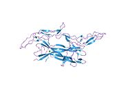
Low-affinity nerve growth factor receptor
Ensembl | |||||||||
|---|---|---|---|---|---|---|---|---|---|
| UniProt | |||||||||
| RefSeq (mRNA) | |||||||||
| RefSeq (protein) | |||||||||
| Location (UCSC) | Chr 17: 49.5 – 49.52 Mb | Chr 11: 95.46 – 95.48 Mb | |||||||
| PubMed search | [3] | [4] | |||||||
| View/Edit Human | View/Edit Mouse |
The p75 neurotrophin receptor (p75NTR) was first identified in 1973 as the low-affinity nerve growth factor receptor (LNGFR)
Receptor family
p75NTR is a member of the tumor necrosis factor receptor superfamily. p75NTR/LNGFR was the first member of this large family of receptors to be characterized,[5][6][11] that now contains about 25 receptors, including tumor necrosis factor 1 (TNFR1) and TNFR2, Fas, RANK, and CD40. All members of the TNFR superfamily contain structurally related cysteine-rich modules in their ECDs. p75NTR is an unusual member of this family due to its propensity to dimerize rather than trimerize, because of its ability to act as a tyrosine kinase co-receptor, and because the neurotrophins are structurally unrelated to the ligands, which typically bind TNFR family members. Indeed, with the exception of p75NTR, essentially all members of the TNFR family preferentially bind structurally related trimeric Type II transmembrane ligands, members of the TNF ligand superfamily.[12]
Structure
p75NTR is a
The p75ECD-binding interface to
Function
Interactions with neurotrophins
Interactions with proneurotrophins
Proforms of NGF and BDNF (proNGF and proBDNF) are precursors to NGF and BDNF. proNGF and proBDNF interact with p75NTR and cause p75NTR-mediated apoptosis without activating TrkA-mediated survival mechanisms. Cleavage of proforms into mature Neurotrophins allows the mature NGF and BDNF to activate TrkA-mediated survival mechanisms.[18][19]
Sensory development
Recent research has suggested a number of roles for the LNGFR, including in development of the eyes and sensory neurons,
Interactions with other receptors
Sortilin
Sortilin is required for many apoptosis-promoting p75NTR reactions, functioning as a co-receptor for the binding of neurotrophins such as BDNF. pro-neurotrophins (such as proBDNF) bind especially well to p75NTR when sortilin is present.[26]
Crosstalk with Trk receptors
When p75NTR initiates apoptosis,
Nogo-66 receptor (NgR1)
p75NTR functions in a complex with Nogo-66 receptor (NgR1) to mediate RhoA-dependent inhibition of growth of regenerating axons exposed to inhibitory proteins of CNS myelin, such as Nogo, MAG or OMgP. Without p75NTR, OMgP can activate RhoA and inhibit CNS axon regeneration. Coexpression of p75NTR and OMgP suppress RhoA activation. A complex of NgR1, p75NTR and LINGO1 can activate RhoA.[27]
p75NTR-mediated signaling pathways
NF-kB activation
NF-kB activity is activated by p75NTR, and is not activated via Trk receptors. NF-kB activity does not effect Brain-derived neurotrophic factor promotion of neuronal survival.[17]
RhoGDI and RhoA
p75NTR serves as a regulator for actin assembly. Ras homolog family member A (RhoA) causes the actin cytoskeleton to become rigid which limits growth cone mobility and inhibits neuronal elongation in the developing nervous system. p75NTR without a ligand bound activates RhoA and limits actin assembly, but neurotrophin binding to p75NTR can inactivate RhoA and promote actin assembly.[28] p75NTR associates with the Rho GDP dissociation inhibitor (RhoGDI), and RhoGDI associates with RhoA. Interactions with Nogo can strengthen the association between p75NTR and RhoGDI. Neurotrophin binding to p75NTR inhibits the association of RhoGDI and p75NTR, thereby suppressing RhoA release and promoting growth cone elongation (inhibiting RhoA actin suppression).[29]
JNK signaling pathway
JNK-Bim-EL signaling pathway
JNK can directly phosphorylate Bim-EL, a splicing
Caspase-dependent signaling
LNGFR also activates a caspase-dependent signaling pathway that promotes developmental axon pruning, and axon degeneration in neurodegenerative disease.[31]
In the apoptosis pathway, members of the TNF receptor superfamily assemble a death-inducing signaling complex (DISC) in which TRADD or FADD bind directly to the receptor's death domain, thereby allowing aggregation and activation of Caspase 8 and subsequent activation of the Caspase cascade. However, Caspase 8 induction does not appear to be involved in p75NTR-mediated apoptosis, but Caspase 9 is activated during p75NTR-mediated killing.[12]
Role in disease
Huntington's disease
Huntington's disease is characterized by cognitive impairments. There is increased expression of p75NTR in the hippocampus of Huntington's disease patients (including mice models and humans). Over expression of p75NTR in mice causes cognitive impairments similar to Huntington's disease. p75NTR is linked to reduced numbers of dendritic spines in the hippocampus, likely through p75NTR interactions with Transforming protein RhoA. Modulating p75NTR function could be a future direction in treating Huntington's disease.[32]
Amyotrophic lateral sclerosis
Amyotrophic lateral sclerosis ALS is a neurodegenerative disease characterized by progressive muscular paralysis reflecting degeneration of motor neurons in the primary motor cortex, corticospinal tracts, brainstem and spinal cord. One study using the superoxide dismutase 1 (SOD1) mutant mouse, an ALS model which develops severe neurodegeneration, the expression of p75NTR correlated with the extent of degeneration and p75NTR knockdown delayed disease progression.[33][34][35]
Alzheimer's disease
Alzheimer's disease (AD) is the most common cause of dementia in the elderly. AD is a neurodegenerative disease characterized by the loss of cognitive functioning - thinking, remembering and reasoning- and behavioral abilities to such an extent that it interferes with a person's daily life and activities. The neuropathological hallmarks of AD include amyloid plaques and neurofibrillary tangles, which lead to neuronal death. Studies in animal models of AD have shown that p75NTR contributes to amyloid β-induced neuronal damage.[36] In humans with AD, increases in p75NTR expression relative to TrkA have been suggested to be responsible for the loss of cholinergic neurons.[37][38] Increases in proNGF in AD [39] indicate that the Neurotrophin environment is favorable for p75NTR/sortilin signaling and supports the theory that age-related neural damage is facilitated by a shift toward proNGF-mediated signaling.[35] A recent study found that activation of Ngfr signaling in astroglia of Alzheimer's disease mouse model enhanced neurogenesis and reduced two hallmarks of Alzheimer's disease.[40] This study also found that NGFR signaling in humans is age-related and correlates with proliferative potential of neural progenitors.
Role in cancer stem cells
p75NTR has been implicated as a marker for cancer stem cells in melanoma and other cancers. Melanoma cells transplanted into an immunodeficient mouse model were shown to require expression of CD271 in order to grow a melanoma.[41] Gene knockdown of CD271 has also been shown to abolish neural crest stem cell properties of melanoma cells and decrease genomic stability leading to a reduced migration, tumorigenicity, proliferation and induction of apoptosis.[42][43][44] Furthermore, increased levels of CD271 were observed in brain metastatic melanoma cells whereas resistance to the BRAF inhibitor vemurafenib supposedly selects for highly malignant brain and lung-metastasizing melanoma cells.[45][44][46][47] Recently, expression of p75NTR (NGFR) was associated with progressive intracranial disease in melanoma patients [48]
Interactions
Low-affinity nerve growth factor receptor has been shown to
References
- ^ a b c GRCh38: Ensembl release 89: ENSG00000064300 – Ensembl, May 2017
- ^ a b c GRCm38: Ensembl release 89: ENSMUSG00000000120 – Ensembl, May 2017
- ^ "Human PubMed Reference:". National Center for Biotechnology Information, U.S. National Library of Medicine.
- ^ "Mouse PubMed Reference:". National Center for Biotechnology Information, U.S. National Library of Medicine.
- ^ S2CID 22472119.
- ^ S2CID 4342838.
- ^ PMID 9927421.
- S2CID 38060583.
- S2CID 15080734.
- S2CID 17364992.
- S2CID 46060440.
- ^ S2CID 8659757.
- ^ PMID 17681869.
- S2CID 40720230.
- PMID 28215307.
- ^ PMID 15470142.
- ^ S2CID 25648122.
- ^ S2CID 872149.
- PMID 20036257.
- PMID 18958367.
- PMID 19210757.
- PMID 19128208.
- PMID 19429149.
- PMID 19553472.
- S2CID 31230806.
- ^ PMID 15987945.
- ^ S2CID 2344794.
- S2CID 17271817.
- ^ S2CID 10865814.
- ^ PMID 9852160.
- PMID 23223278.
- PMID 25180603.
- PMID 24475283.
- S2CID 5901529.
- ^ PMID 26109945.
- PMID 18978948.
- PMID 16619032.
- S2CID 38106502.
- S2CID 8443739.
- PMID 37429840.
- PMID 20596026.
- PMID 24799129.
- PMID 28112719.
- ^ PMID 28852061.
- PMID 25674270.
- PMID 25725450.
- PMID 30053879.
- PMID 36435874.
- S2CID 13116876.
- ^ PMID 12414813.
- PMID 14593116.
- PMID 12716928.
- PMID 10764727.
- S2CID 4343450.
- PMID 12682012.
- ^ PMID 10514511.
- PMID 9626059.
Further reading
- Buxser S, Puma P, Johnson GL (February 1985). "Properties of the nerve growth factor receptor. Relationship between receptor structure and affinity". The Journal of Biological Chemistry. 260 (3): 1917–1926. PMID 2981877.
- Glass DJ, Nye SH, Hantzopoulos P, Macchi MJ, Squinto SP, Goldfarb M, Yancopoulos GD (July 1991). "TrkB mediates BDNF/NT-3-dependent survival and proliferation in fibroblasts lacking the low affinity NGF receptor". Cell. 66 (2): 405–413. S2CID 43626580.
- Ibáñez CF (June 2002). "Jekyll-Hyde neurotrophins: the story of proNGF". Trends in Neurosciences. 25 (6): 284–286. S2CID 9449831.
- Radeke MJ, Misko TP, Hsu C, Herzenberg LA, Shooter EM (1987). "Gene transfer and molecular cloning of the rat nerve growth factor receptor". Nature. 325 (6105): 593–597. S2CID 4342838.
External links
- Nerve+Growth+Factor+Receptor,+Low-Affinity at the U.S. National Library of Medicine Medical Subject Headings (MeSH)

