
Lumbar vertebrae
| Lumbar vertebrae | |
|---|---|
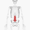
 Position of human lumbar vertebrae (shown in red). It consists of 5 bones, from the top down, L1, L2, L3, L4 and L5. | |
 Animation of L1 | |
| Details | |
| Insertions | Psoas major muscle |
| Nerve | Lumbar nerves |
| Identifiers | |
| Latin | vertebrae lumbales |
| MeSH | D008159 |
| TA98 | A02.2.04.001 |
| TA2 | 1068 |
| FMA | 9921 |
| Anatomical terms of bone | |
The lumbar vertebrae are located between the
Human anatomy
In human anatomy, the five
General characteristics

The adjacent figure depicts the general characteristics of the first through fourth lumbar vertebrae. The fifth vertebra contains certain peculiarities, which are detailed below.
As with other vertebrae, each lumbar vertebra consists of a vertebral body and a vertebral arch. The vertebral arch, consisting of a pair of pedicles and a pair of laminae, encloses the vertebral foramen (opening) and supports seven processes.
Body
The
Arch
The
The
The vertebral foramen within the arch is triangular, larger than the thoracic vertebrae, but smaller than in the cervical vertebrae.[1]
Processes
The
The superior and inferior articular processes are well-defined, projecting respectively upward and downward from the junctions of pedicles and laminae. The facets on the superior processes are concave, and look backward and medialward; those on the inferior are convex, and are directed forward and lateralward. The former are wider apart than the latter since in the articulated column, the inferior articular processes are embraced by the superior processes of the subjacent vertebra.[1]
The
Three portions or tubercles can be noticed in a transverse process of a lower lumbar vertebrae: the lateral or
First and fifth lumbar vertebrae

The first lumbar vertebra is level with the anterior end of the
The fifth lumbar vertebra is characterized by its body being much deeper in front than behind, which accords with the prominence of the sacrovertebral articulation; by the smaller size of its spinous process; by the wide interval between the inferior articular processes, and by the thickness of its transverse processes, which spring from the body as well as from the pedicles.[1] The fifth lumbar vertebra is by far the most common site of spondylolysis and spondylolisthesis.[3]
Most individuals have five lumbar vertebrae, while some have four or six. Lumbar disorders that normally affect L5 will affect L4 or L6 in these latter individuals.
Segmental movements
The range of segmental movements in a single segment is difficult to measure clinically, not only because of variations between individuals, but also because it is age and sex dependent. Furthermore, flexion and extension in the lumbal spine is the product of a combination of rotation and translation in the sagittal plane between each vertebra.[4]
Ranges of segmental movements in the lumbar spine (White and Punjabi, 1990) are (in degrees): [5]
| L1-L2 | L2-L3 | L3-L4 | L4-L5 | L5-S1 | |
|---|---|---|---|---|---|
| Flexion/ Extension |
12° | 14° | 15° | 16° | 17° |
| Lateral flexion |
6° | 6° | 8° | 6° | 3° |
| Axial rotation |
2° | 2° | 2° | 2° | 1° |
Congenital anomalies
Congenital vertebral anomalies can cause compression of the spinal cord by deforming the vertebral canal or causing instability.
-
 Lumbarization ofribs.
 -
 Sacralization of the L5 vertebra is seen at the lower right of the image.
 -
 Congenital block vertebra of the lumbar spine. CT volume rendering.


Other apes
African apes have three and four lumbar vertebrae, (bonobos have longer spines with an additional vertebra) and humans normally five. This difference, and because the lumbar spines of the extinct Nacholapithecus (a Miocene hominoid with six lumbar vertebrae and no tail) are similar to those of early Australopithecus and early Homo, it is assumed that the chimpanzee–human last common ancestor also had a long vertebral column with a long lumbar region and that the reduction in the number of lumbar vertebrae evolved independently in each ape clade.[6] The limited number of lumbar vertebrae in chimpanzees and gorillas result in an inability to lordose (curve) their lumbar spines, in contrast to the spines of Old World monkeys and Nacholapithecus and Proconsul, which suggests that the last common ancestor was not "short-backed" as previously believed. [7]
Additional images
MRI
-
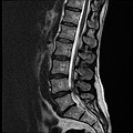 MRI lumbar spine with degeneration (sagittal T2 FRFSE)
 -
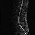
 MRI lumbar spine with degeneration (sagittal T1 FSE)
 -
 MRI lumbar spine with degeneration (sagittal FAST STIR)
 -
MRI lumbar spine pre-hemilaminectomy (sagittal T2 FRFSE)
-
MRI lumbar spine pre-hemilaminectomy (sagittal T1 FSE)
-
MRI lumbar spine pre-hemilaminectomy (sagittal FAST STIR)
-
MRI lumbar spine post-hemilaminectomy (sagittal T2 FRFSE)
-
MRI lumbar spine post-hemilaminectomy (sagittal T1 FSE)
-
Contrast MRI lumbar spine post-hemilaminectomy (sagittal T1 FSE FS)


Illustrations
-
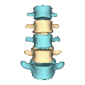
 3D image of a lumbar vertebra
 -
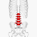
 Position of lumbar vertebrae (shown in red). Animation.
 -
 Same as the left. Bones around the lumbar vertebrae are shown as semi-transparent.
 -
 Shape of lumbar vertebrae (shown in blue and yellow). Animation.
 -
 Vertebral column.
 -
 Muscles of the iliac and anterior femoral regions. First lumbar vertebra second highest vertebra seen.
 -
![Orientation of vertebral column on surface. T3 is at level of medial part of spine of scapula. T7 is at inferior angle of the scapula. L4 is at highest point of iliac crest. S2 is at the level of posterior superior iliac spine. Furthermore, C7 is easily localized as a prominence at the lower part of the neck.[8]](//upload.wikimedia.org/wikipedia/commons/thumb/0/08/Orientation.PNG/108px-Orientation.PNG) Orientation of vertebral column on surface. T3 is at level of medial part ofinferior angle of the scapula. L4 is at highest point of iliac crest. S2 is at the level of posterior superior iliac spine. Furthermore, C7 is easily localized as a prominence at the lower part of the neck.[8]
Orientation of vertebral column on surface. T3 is at level of medial part ofinferior angle of the scapula. L4 is at highest point of iliac crest. S2 is at the level of posterior superior iliac spine. Furthermore, C7 is easily localized as a prominence at the lower part of the neck.[8] -
 Vertebral column
 -
 Illustration highlighting lumbar spine.
 -
 A lumbar vertebra seen from the side
 -
 Ossification of lumbar vertebrae


![Orientation of vertebral column on surface. T3 is at level of medial part of spine of scapula. T7 is at inferior angle of the scapula. L4 is at highest point of iliac crest. S2 is at the level of posterior superior iliac spine. Furthermore, C7 is easily localized as a prominence at the lower part of the neck.[8]](/File:Orientation.PNG)


See also
References
![]() This article incorporates text in the public domain from page 104 of the 20th edition of Gray's Anatomy (1918)
This article incorporates text in the public domain from page 104 of the 20th edition of Gray's Anatomy (1918)
- ^ a b c d e f g h Gray's Anatomy (1918), see infobox
- ^ a b Postacchini, Franco (1999) Lumbar Disc Herniation p.19
- ^ Eizenberg, N. et al. (2008). General Anatomy: Principles and Applications, p. 17.
- S2CID 43352264. Archived from the originalon November 14, 2010.
- ^ Hansen; et al. (2006). "Ranges of Segmental Motion for the Lumbar Spine". Medscape. Archived from the original on July 5, 2015.
- PMID 19688850. (Abstract)
- PMID 20855303. (Introduction)
- ^ Anatomy Compendium (Godfried Roomans and Anca Dragomir)
External links
- "Lower Back Pain Condition, Treatment and Exercise". SpineUniverse. Retrieved February 15, 2017.
- "Virtual Spine — Online Learning Resource". Toronto Western Hospital Department of Anesthesia and Pain Management. Retrieved February 15, 2017.
| National | |
|---|---|
| Other | |