
Lunate bone
| Lunate bone | |
|---|---|
| Identifiers | |
| Latin | os lunatum |
| MeSH | D012667 |
| TA98 | A02.4.08.005 |
| TA2 | 1252 |
| FMA | 23712 |
| Anatomical terms of bone] | |
The lunate bone (semilunar bone) is a
Structure
The lunate is a crescent-shaped
The lunate is stabilised by a medial ligament to the scaphoid bone and a lateral ligament to the triquetral bone. Ligaments between the radius and carpal bone also stabilise the position of the lunate, as does its position in the lunate fossa of the radius.[3]
Bone
The proximal surface of the lunate bone is smooth and convex, articulating with the radius. The lateral surface is flat and narrow, with a crescentic facet for articulation with the scaphoid bone. The medial surface possesses a smooth and quadrilateral facet for articulation with the triquetral bone. The palmar surface is rough, as is the dorsal surface. The dorsal surface is broad and rounded. The distal surface of the bone is deep and concave.[4]
Blood supply
The lunate receives its blood supply from dorsal and palmar branches.[3]
Variation
The lunate has a variable shape. About one-third of lunate bones do not possess a medial facet, meaning they do not articulate with the hamate bone. Additionally, in about 20% of people, blood supply may arise from palmar vessels alone.[3]
Ossification
The ossification of the lunate bone commences between 18 months and 4 years 3 months.[5]
Function
The carpal bones function as a unit to provide a bony superstructure for the hand.[2]: 708 As a proximal carpal bone, the lunate is also involved in movement of the wrist.[3]
Clinical relevance

The lunate bone is the most frequently dislocated carpal bone.
- Carpal coalition
- Kienbock's disease
- Teisen classification
Etymology
The name of the lunate bone derives from the "crescent-shaped" (
luna ("moon"), from the bone's resemblance to a crescent moon. In amphibians and reptiles, the bone is instead referred to as the intermedium, because of its position between the other two proximal carpals.Additional images
-
 Lunate bone of the left hand (shown in red). Animation.
 -
 Lunate bone of the left hand. Close up. Animation.
 -
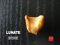 Lunate bone.
 -
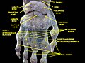
 Bones of the left hand. Volar surface.
 -
 Bones of the left hand. Dorsal surface.
 -
 Cross section of wrist (thumb on left). Lunate shown in red.
 -
 Dislocated lunate
 -
 Dislocated lunate
 -
 Wrist joint. Deep dissection. Posterior view.
 -
 Wrist joint. Deep dissection. Posterior view.
_-_animation01.gif)
_-_animation02.gif)


See also
- Carpal bone
References
![]() This article incorporates text in the public domain from page 224 of the 20th edition of Gray's Anatomy (1918)
This article incorporates text in the public domain from page 224 of the 20th edition of Gray's Anatomy (1918)
- ^ Manaster, B. J., Julia Crim "Imaging Anatomy: Musculoskeletal E-Book" Elsevier Health Sciences, 2016, p. 326.
- ^ ISBN 978-0-8089-2306-0.
- ^ PMID 15831311.
- ^ Gray, Henry (1918). Anatomy of the Human Body. p. 6b. The Hand. 1. The Carpus. Retrieved 5 January 2014.
- ISSN 0973-9130. Retrieved 18 August 2014.
- ^ Harper, Douglas. "Lunate". Online Etymology Dictionary. Retrieved 5 January 2014.
| National | |
|---|---|
| Other | |