
Lymphedema
| Lymphedema | |
|---|---|
| Other names | Lymphoedema, lymphatic obstruction, lymphatic insufficiency |
Based on symptoms[2] | |
| Differential diagnosis | Lipodystrophy, venous insufficiency[2] |
Lymphedema, also known as lymphoedema and lymphatic edema, is a condition of localized
Lymphedema is most frequently a complication of cancer treatment or
While there is no cure, treatment may improve outcomes.
Signs and symptoms

The most common manifestation of lymphedema is soft tissue swelling (edema). As the disorder progresses, worsening edema and skin changes including discoloration, verrucous (wart-like) hyperplasia, hyperkeratosis, papillomatosis, dermal thickening, and ulcers may be seen. Additionally, there is increased risk of infection of the skin, known as erysipelas.[citation needed]
Complications
When lymphatic impairment becomes so great that the collected lymph fluid exceeds the lymphatic system's ability to transport it, an abnormal amount of protein-rich fluid collects in the tissues. Left untreated, this stagnant, protein-rich fluid causes tissue channels to increase in size and number, reducing oxygen availability. This interferes with wound healing and provides a rich medium for bacterial growth which can result in
In rare cases, lymphedema may lead to a form of cancer called
Lymphedema can be disfiguring, and may result in a poor body image and psychological distress.[8] Complications of lymphedema can cause difficulties in activities of daily living.[9]
Causes and risk factors
Lymphedema may be inherited (primary) or caused by injury to the lymphatic vessels (secondary).[10] There are also risk factors that may increase one's risk of developing lymphedema such as old age, being overweight or obese, and having rheumatic or psoriatic arthritis.[11]
Lymph node damage
Lymphedema is most commonly seen after lymph node dissection, surgery or radiation therapy for the treatment of cancer, most notably breast cancer. In many patients the condition does not develop until months or even years after therapy has concluded.[medical citation needed] Lymphedema may also be associated with accidents or certain diseases or conditions that may inhibit the lymphatic system from functioning properly.[4] It can also be caused by damage to the lymphatic system from infections such as cellulitis.[12] In tropical areas of the world where parasitic filarial worms are endemic, a common cause of secondary lymphedema is filariasis.[13]
Primary lymphedema may be congenital or may arise sporadically. Multiple syndromes are associated with primary lymphedema, including Turner syndrome, Milroy's disease, and Klippel–Trénaunay syndrome. In these syndromes it may occur as a result of absent or malformed lymph nodes and/or lymphatic channels. Lymphedema can be present at birth, develop at the onset of puberty (praecox), or not become apparent for many years into adulthood (tarda). In men, lower-limb primary lymphedema is most common, occurring in one or both legs. Some cases of lymphedema may be associated with other vascular abnormalities.[4][citation needed]
Secondary lymphedema affects both men and women, and, in Western countries, is most commonly due to cancer treatment.
Head and neck lymphedema can be caused by surgery or radiation therapy for tongue or throat cancer. It may also occur in the lower limbs or groin after surgery for colon, ovarian or uterine cancer, if removal of lymph nodes or radiation therapy is required. Surgery or treatment for prostate, colon and testicular cancers may result in secondary lymphedema, particularly when lymph nodes have been removed or damaged.[medical citation needed]
The onset of secondary lymphedema in patients who have had cancer surgery has also been linked to aircraft flight (likely due to decreased cabin pressure or relative immobility). For cancer survivors wearing a prescribed and properly fitted compression garment may help decrease swelling during air travel.[22]
Some cases of lower-limb lymphedema have been associated with the use of tamoxifen, due to blood clots and deep vein thrombosis (DVT) associated with this medication. Resolution of the blood clots or DVT is needed before lymphedema treatment can be initiated.[medical citation needed]
At birth
Hereditary lymphedema is a primary lymphedema – swelling that results from abnormalities in the
The most common cause is
One defined genetic cause for hereditary lymphedema is
Primary lymphedema occurs in approximately 1-3 births out of every 10,000 births, with a female to male ratio of 3.5:1. In North America, the incidence of primary lymphedema is approximately 1.15 births out of every 100,000 births.[contradictory] Compared to secondary lymphedema, primary lymphedema is relatively rare.[27]
Inflammatory lymphedema
Physiology
Lymph is formed from the fluid that filters out of blood and contains proteins, cellular debris, bacteria, etc. This fluid is collected by the initial lymph collectors that are blind-ended endothelial-lined vessels with fenestrated openings that allow fluids and particles as large as cells to enter. Once inside the lumen of the lymphatic vessels, the fluid is guided along increasingly larger vessels, first with rudimentary valves to prevent backflow, later with complete valves similar to the venous valve. Once the lymph enters the fully valved lymphatic vessels, it is pumped by a rhythmic peristaltic-like action by smooth muscle cells within the lymphatic vessel walls. This peristaltic action is the primary driving force moving lymph within its vessel walls. The sympathetic nervous system regulates of the frequency and power of the contractions. Lymph movement can be influenced by the pressure of nearby muscle contraction, arterial pulse pressure and the vacuum created in the chest cavity during respiration, but these passive forces contribute only a minor percentage of lymph transport. The fluids collected are pumped into continually larger vessels and through lymph nodes, which remove debris and police the fluid for dangerous microbes. The lymph ends its journey in the thoracic duct or right lymphatic duct, which drain into the blood circulation.[10]
Diagnosis
Diagnosis is generally based on signs and symptoms, with testing used to rule out other potential causes.[2] An accurate diagnosis and staging may help with management.[2] A swollen limb can result from different conditions that require different treatments. Diagnosis of lymphedema is currently based on history, physical exam, and limb measurements. Imaging studies such as lymphoscintigraphy and indocyanine green lymphography are only required when surgery is being considered.[2] However, the ideal method of staging to guide treatment is controversial because of several different proposed protocols.[30][31]
Lymphedema can occur in both the upper and lower extremities, and in some cases, the head and neck. Assessment of the extremities first begins with a visual inspection; color, presence of hair, visible veins, size and any sores or ulcerations are noted. Lack of hair may indicate an arterial circulation problem.[32] In cases of swelling, the extremities' circumference is measured over time for reference. In early stages of lymphedema, elevating the limb may reduce or eliminate the swelling. Palpation of the wrist or ankle can determine the degree of swelling; assessment includes a check of the pulses. The axillary or inguinal lymph nodes may be enlarged due to the swelling. Enlargement of the nodes lasting more than three weeks may indicate infection or other illnesses (such as sequela from breast cancer surgery) requiring further medical attention.[32]
Diagnosis or early detection of lymphedema is difficult. The first signs may be subjective observations such as a feeling of heaviness in the affected extremity. These may be symptomatic of early-stage lymphedema where accumulation of lymph is mild and not detectable by changes in volume or circumference. As lymphedema progresses, definitive diagnosis is commonly based upon an objective measurement of differences between the affected or at-risk limb and the opposite unaffected limb, e.g. in volume or circumference. No generally accepted criterion is definitively diagnostic, although a volume difference of 200 ml between limbs or a 4 cm (1.6 in) difference (at a single measurement site or set intervals along the limb) is often used. Bioimpedance measurement (which measures the amount of fluid in a limb) offers greater sensitivity than other methods.[33] Devices like SOZO [34] utilize Bioimpedence Analysis (BIA) by sending a current through the body and measuring the resultant impedance. Another approach involves Tissue Dielectric Constant (TDC) measurement, used by devices such as Delfin Technology's MoistureMeterD and LymphScanner [35], which employ microwaves to detect changes in the dielectric properties of tissue. These innovative techniques have become integral to official protocols for lymphedema detection [36].
Chronic venous stasis changes can mimic early lymphedema, but are more often bilateral and symmetric. Lipedema can also mimic lymphedema, however lipedema characteristically spares the feet beginning abruptly at the malleolus (ankle).[2] As a part of the initial work-up before diagnosing lymphedema, it may be necessary to exclude other potential causes of lower extremity swelling such as kidney failure, hypoalbuminemia, congestive heart-failure, protein-losing kidney disease, pulmonary hypertension, obesity, pregnancy and drug-induced edema.[citation needed]
Classification

The International Society of Lymphology (ISL) Staging System is based solely on subjective symptoms, making it prone to substantial observer bias. Imaging modalities have been suggested as useful adjuncts to the ISL staging to clarify the diagnosis, such as Cheng's Lymphedema Grading tool, which assesses the severity of extremity lymphedema based on objective limb measurements and provides appropriate options for management.[37][38][39]
I. Grading

- Grade 1: Spontaneously reversible on elevation. Mostly pitting edema.
- Grade 2: Non-spontaneously reversible on elevation. Mostly non-pitting edema.
- Grade 3: Gross increase in volume and circumference of Grade 2 lymphedema, with eight stages of severity given below based on clinical assessments.
II. Staging
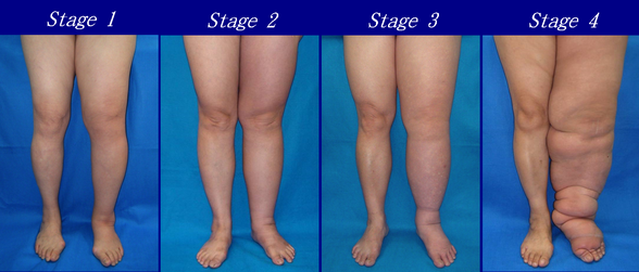
As described by the Fifth
- Stage 0: The lymphatic vessels have sustained some damage that is not yet apparent. Transport capacity is sufficient for the amount of lymph being removed. Lymphedema is not present.
- Stage 1 : Swelling increases during the day and disappears overnight as the patient lies flat in bed. Tissue is still at the pitting stage: when pressed by the fingertips, the affected area indents and reverses with elevation. Usually, upon waking in the morning, the limb or affected area is normal or almost normal in size. Treatment is not necessarily required at this point.
- Stage 2: Swelling is not reversible overnight, and does not disappear without proper management. The tissue now has a spongy consistency and is considered non-pitting: when pressed by the fingertips, the affected area bounces back without indentation. Fibrosis found in Stage 2 lymphedema marks the beginning of the hardening of the limbs and increasing size.
- Stage 3: Swelling is irreversible and usually the limb(s) or affected area becomes increasingly large. The tissue is hard (fibrotic) and unresponsive; some patients consider undergoing reconstructive surgery, called "debulking". This remains controversial, however, since the risks may outweigh the benefits and further damage done to the lymphatic system may make the lymphedema worse.
- Stage 4: The size and circumference of the affected limb(s) become noticeably larger. Bumps, lumps, or protrusions (also called knobs) on the skin begin to appear.
- Stage 5: The affected limb(s) become grossly large; one or more deep skin folds is present.
- Stage 6: Knobs of small elongated or rounded sizes cluster together, giving mossy-like shapes on the limb. Mobility of the patient becomes increasingly impaired.
- Stage 7: The person becomes "handicapped", and is unable to independently perform daily routine activities such as walking, bathing and cooking. Assistance from the family and health care system is needed.
Grades
Lymphedema can also be categorized by its severity (usually compared to a healthy extremity):[44]
- Grade 1 (mild edema): Involves the distal parts such as a forearm and hand or a lower leg and foot. The difference in circumference is less than 4 cm (1.6 in) and no other tissue changes are present.
- Grade 2 (moderate edema): Involves an entire limb or corresponding quadrant of the trunk. Difference in circumference is 4–6 cm (1.6–2.4 in). Tissue changes, such as pitting, are apparent. The patient may experience erysipelas.
- Grade 3a (severe edema): Lymphedema is present in one limb and its associated trunk quadrant. Circumferential difference is greater than 6 cm (2.4 in). Significant skin alterations, such as fistulae, are present. Additionally, the patient may experience repeated attacks of erysipelas.
- Grade 3b (massive edema): The same symptoms as grade 3a, except that two or more extremities are affected.
- Grade 4 (gigantic edema): In this stage of lymphedema, the affected extremities are huge, due to almost complete blockage of the lymph channels.
Differential
Lymphedema should not be confused with edema arising from chronic venous insufficiency, which is caused by compromise of venous drainage rather than lymphatic drainage. However, untreated venous insufficiency can progress into a combined venous/lymphatic disorder known as phlebetic lymphedema (or phlebolymphedema).[45][46][47]
Treatment
While there is no cure, treatment may improve outcomes.[2] This commonly include compression therapy, good skin care, exercise, manual lymphatic drainage (MLD) and the use of an intermittent pneumatic compression pump, which together is known as combined decongestive therapy.[2] MLD is most effective in mild to moderate disease.[48] In breast cancer-related lymphedema, MLD is safe and may offer added benefit to compression bandages for reducing swelling.[48] Most people with lymphedema can be medically managed with conservative treatment.[49] Diuretics are not useful.[2] Surgery is generally only used if symptoms are not improved by other measures.[2]
Compression
Garments

Once a person is diagnosed with lymphedema, compression becomes imperative in the management of the condition. Garments are often intended to be worn all day but may be taken off for sleep, unless otherwise prescribed. Elastic compression garments are worn on the affected limb following complete de-congestive therapy to maintain edema reduction. Inelastic garments provide containment and reduction.[2] Available styles, options, and prices vary widely. A professional garment fitter or certified lymphedema therapist can help determine the best option for the patient.[citation needed]
Bandaging
Compression bandaging, also called wrapping, is the application of layers of padding and short-stretch bandages to the involved areas. Short-stretch bandages are preferred over long-stretch bandages (such as those normally used to treat sprains), as the long-stretch bandages cannot produce the proper therapeutic tension necessary to safely reduce lymphedema and may produce a tourniquet effect. Compression bandages provide resistance that assists in pumping fluid out of the affected area during exercise. This counter-force results in increased lymphatic drainage and therefore a decrease in size of the swollen area.[50]
Intermittent pneumatic compression therapy
Intermittent pneumatic compression therapy (IPC) utilizes a multi-chambered pneumatic sleeve with overlapping cells to promote movement of lymph fluid.[2] Pump therapy should only be used in addition to other treatments such as compression bandaging and manual lymph drainage. Pump therapy has been used in the past to help with controlling lymphedema. In some cases, pump therapy helps soften fibrotic tissue and therefore potentially enable more efficient lymphatic drainage.[51] However, reports link pump therapy to increased incidence of edema proximal to the affected limb, such as genital edema arising after pump therapy in the lower limb.[52] Current literature has suggested the use of IPC treatment in conjunction with an elastic therapeutic tape is more effective in the overall reduction of lymphedema as well as increasing shoulder range of motion than the traditional treatment of IPC paired with complete decongestive therapy. The tape is an elastic cotton strip with an acrylic adhesive that is used commonly used to relieve the discomfort and disability associated with sports injuries, but in the context of lymphedema, this increases the space between the dermis and the muscle which increases the opportunity for lymphatic fluid to flow out naturally.[53] The use of IPC treatments with tape, as well as subsequent lymphatic drainage, has proven to significantly reduce the circumference of lymphatic limbs in patients experiencing lymphedema secondary to breast cancer post-mastectomy.[53]
Exercise
In those with lymphedema or at risk of developing lymphedema, such as following breast cancer treatment, resistance training did not increase swelling and led to decreases in some, in addition to other potential beneficial effects on cardiovascular health.[54][55] Moreover, resistance training and other forms of exercise were not associated with an increased risk of developing lymphedema in people who previously received breast cancer-related treatment. Compression garments should be worn during exercise.[56]
Physical therapy for patients with lymphedema may include trigger point release, soft tissue massage, postural improvement, patient education on condition management, strengthening, and stretching exercises. Exercises may increase in intensity and difficulty over time, beginning with passive movements to increase range of motion and progressing towards using external weights and resistance in various postures.[50]
Surgery
The treatment of lymphedema is usually conservative, however the use of surgery is proposed for some cases.[57]
Suction assisted lipectomy (SAL), also known as liposuction for lymphedema, may help improve chronic non pitting edema.[58] The procedure removes fat and protein and is done alongside continued compression therapy.[58]
Vascularized lymph node transfers (VLNT) and lymphovenous bypass are supported by tentative evidence as of 2017[update] but are associated with a number of complications.
Laser therapy
Epidemiology
Lymphedema affects approximately 200 million people worldwide.[4]
References
- PMID 8342046.
- ^ PMID 29132859.
- PMID 30725924.
- ^ PMID 29132848.
- PMID 6541442.
- PMID 18065640.
- PMID 9486670.
- ^ Publishing L (2009-10-28). "Body image and quality of life in secondary lymphedema of the upper limb". Servier – Phlebolymphology. Archived from the original on 2019-09-03. Retrieved 2019-09-03.
- ^ "Oncology Fact Sheet" (PDF). aota.org. Retrieved 2019-09-03.
- ^ a b "A Visual Guide to Lymphedema". WebMD. Retrieved 7 July 2021.
- ^ "Lymphedema – Symptoms and causes". Mayo Clinic. Retrieved 2023-08-02.
- ^ "Lymphoedema – Causes". nhs.uk. 2017-10-20. Retrieved 2022-06-15.
- PMID 18830049.
- ^ PMID 18720912.
- ISBN 978-0-89793-458-9.
- ^ Choices N. "IPS retired". nhs.uk. Archived from the original (PDF) on 28 August 2021. Retrieved 9 May 2018.
- S2CID 25777064.
- PMID 1411352.
- PMID 7899840.
- S2CID 36177502.
- PMID 6386761.
- PMID 24591279.
- ^ Liem TK, Moneta GL (2010). "Chapter 24. Venous and Lymphatic Disease". In Brunicardi FC, Andersen DK, Billiar TR, Dunn DL, Hunter JG, Matthews JB, Pollock RE (eds.). Schwartz's Principles of Surgery (9th ed.). New York, NY: The McGraw-Hill Companies.[permanent dead link]
- ^ Burkhart CN, Adigun C, Burton CS (2012). "Chapter 174. Cutaneous Changes in Peripheral Venous and Lymphatic Insufficiency". In Goldsmith LA, Katz SI, Gilchrest BA, Paller AS, Leffell DJ, Wolff K (eds.). Fitzpatrick's Dermatology in General Medicine (8 ed.). New York, NY: The McGraw-Hill Companies.[permanent dead link]
- PMID 28179280.
- PMID 28643018.
- PMID 7027437.
- PMID 25607253.
- S2CID 6650610.
- S2CID 34921176.
- PMID 12578410.
- ^ ISBN 978-1-4160-5188-6.
- PMID 16569209.
- ^ "SOZO® Digital Health Platform | ImpediMed". www.impedimed.com. 2019-06-05. Retrieved 2024-03-20.
- ^ "Delfin Technologies - Skin research instruments". Delfin Technologies. Retrieved 2024-03-20.
- PMID 36566297.
- PMID 23930436.
- S2CID 13376881.
- ISBN 978-0-323-29897-1.
- ^ ISBN 978-0-323-29897-1. July 2015.
- ^ "Treatment and Prevention of Problems Associated with Lymphatic Filariasis" (PDF). World Health Organization. Archived (PDF) from the original on 2012-04-18. Retrieved 2014-05-16.
- PMID 1441569.
- ISBN 978-1-84628-793-0.[page needed]
- PMID 28453410.
- PMID 21326689.
- PMID 24527138.
- ^ Publishing L, Guiboles (2009-11-24). "The causes of edema in chronic venous insufficiency". Servier – Phlebolymphology. Retrieved 2023-09-01.
- ^ PMID 25994425.
- PMID 17175115.
- ^ PMID 31367497.
- PMID 12903017.
- PMID 9561507.
- ^ S2CID 3351415.
- PMID 27650122.
- S2CID 3125299.
- ^ "Position Paper: Exercise | National Lymphedema Network". Lymphnet.org. Archived from the original on 2014-05-08. Retrieved 2014-05-16.
- S2CID 7791985.
- ^ PMID 24558061.
- ^ dotmed.com December 27, 2006 Archived January 7, 2010, at the Wayback Machine Low Level Laser FDA Cleared for the Treatment of Lymphedema. (accessed 9 November 09)
- ^ National Cancer Institute: Low-level laser therapy Archived 2009-09-24 at the Wayback Machine accessed 9 November 09
- S2CID 7043788.