
Magnetic resonance imaging of the brain
| MRI of Brain | |
|---|---|
 Cross-sectional T1-weighted MRI of a healthy human brain acquired with an ultra high-field MR of 7 Tesla field strength | |
| ICD-10-PCS | B030ZZZ |
| ICD-9-CM | 88.91 |
| OPS-301 code | 3-800, 3-820 |
Magnetic resonance imaging of the brain uses
History
The first MR images of a human brain were obtained in 1978 by two groups of researchers at

In the early 1980s to the early 1990s, 'Jedi' helmets, inspired by the 'Return of the Jedi' Star Wars film, were sometimes worn by children in order to obtain good image quality. The copper coils of the helmet were used as a radio aerial to detect the signals while the 'Jedi' association encouraged children to wear the helmets and not be frightened by the procedure. These helmets were no longer needed as MR scanners improved.
In the early 1990s, Peter Basser and Le Bihan, working at
The first study of the human brain at 3.0 T was published in 1994,[13] and in 1998 at 8 T.[14] Studies of the human brain have been performed at 9.4 T (2006)[15] and up to 10.5 T (2019).[16]
Paul Lauterbur and Sir Peter Mansfield were awarded the 2003 Nobel Prize in Physiology or Medicine for their discoveries concerning MRI.

The record for the highest spatial resolution of a whole intact brain (postmortem) is 100 microns, from Massachusetts General Hospital. The data was published in Scientific Data on 30 October 2019.[17][18]
Applications
One advantage of MRI of the brain over
In the case of a concussion, an MRI should be avoided unless there are progressive neurological symptoms, focal neurological findings or concern of skull fracture on exam.[22] In the analysis of a concussion, measurements of Fractional Anisotropy, Mean Diffusivity, Cerebral Blood Flow, and Global Connectivity can be taken to observe the pathophysiological mechanisms being made while in recovery.[23]
In analysis of the fetal brain, MRI provides more information about gyration than ultrasound.[24]
MRI is sensitive for the detection of brain abscess.[25]
A number of different imaging modalities or
- T1-weighted (T1W) images: Cerebrospinal fluid is dark. T1-weighted images are useful for visualizing normal anatomy.
- T2-weighted (T2W) images: CSF is light, but fat (and thus white matter) is darker than with T1. T2-weighted images are useful for visualizing pathology.[26]
- Diffusion-weighted images (DWI): DWI uses the diffusion of water molecules to generate contrast in MR images.
- Proton density (PD) images: CSF has a relatively high level of Gray matter is brighter than white matter.[27]

- demyelination.[29]
Diagnostic Usage
MRI of the brain and head has multiple diagnostic usages, including identifying aneurysms, strokes, tumors and other brain injury.[30] In many diseases, such as Parkinson's or Alzheimer's, MRI is useful to help differentially diagnose against other diseases.[31][32] On the topic of diagnosis, MRI data has been used with deep learning networks to identify brain tumors.[33]
See also
Gallery
-
 Brain regions on T1 MRI
 -
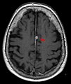 T1 (note CSF is dark) with contrast (arrow pointing to meningioma of the falx)
 -
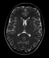 Normal axial T2-weighted MR image of the brain
 -
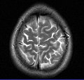 MRI image of the surface of the brain.


References
- ^ "Britain's brains produce first NMR scans". New Scientist: 588. 1978.
- ^ "Blood-flow checker". Popular Science: 12. 1987.
- ^ Le Bihan D, Breton E (1987). "Method to Measure the Molecular Diffusion and/or Perfusion Parameters of Live Tissue". US Patent # 4,809,701.
- S2CID 41228095.
- ISBN 978-1-4419-1328-9. Retrieved 10 June 2015.
- S2CID 36417513.
- S2CID 24795253.
- PMID 19927075.
- ISBN 9780195369779.
- S2CID 42727826.
- PMID 22245338.
- PMID 9205259.
- S2CID 20221062.
- S2CID 41305659.
- PMID 17075852.
- S2CID 208226414.
- ^ "100-Hour-Long MRI of Human Brain Produces Most Detailed 3D Images Yet". 10 July 2019.
- ^ "Team publishes on highest resolution brain MRI scan".
- ISBN 978-3-13-108131-5. Retrieved 18 July 2011.
- ISBN 978-0-7817-2568-2. Retrieved 24 July 2011.
- ISBN 978-0-444-52015-9. Retrieved 18 July 2011.
- ABIM Foundation, American Medical Society for Sports Medicine, retrieved 29 July 2014
- PMID 28280686.)
{{cite journal}}: CS1 maint: multiple names: authors list (link - ISBN 978-3-540-40747-8. Retrieved 24 July 2011.
- PMID 23122258.
- ISBN 978-0-521-81939-8. Retrieved 18 July 2011.
- ISBN 978-0-470-86949-9. Retrieved 18 July 2011.
- ISBN 978-1-4051-9220-0. Retrieved 18 July 2011.
- ISBN 978-0-8493-8138-6. Retrieved 18 July 2011.
- ^ "MRI - Mayo Clinic". www.mayoclinic.org. Retrieved 2023-12-22.
- PMID 28378231.
- PMC 2938772.
- PMID 33094213.
| X-ray/ radiography |
| ||||||||||||
|---|---|---|---|---|---|---|---|---|---|---|---|---|---|
| MRI | |||||||||||||
| Ultrasound | |||||||||||||
| Radionuclide |
| ||||||||||||
| Optical/Laser | |||||||||||||
| Thermography | |||||||||||||
| Target conditions |
| ||||||||||||