
Malleus
| Malleus | |
|---|---|
membrana tympani with the hammer and the chorda tympani, viewed from within, from behind, and from above (malleus visible at center) | |
| Details | |
| Pronunciation | /ˈmæliəs/ |
| Precursor | first branchial arch |
| Part of | Middle ear |
| System | Auditory system |
| Identifiers | |
| Latin | malleus |
| MeSH | D008307 |
| TA98 | A15.3.02.043 |
| TA2 | 881 |
| FMA | 52753 |
| Anatomical terms of bone] | |
 |
| This article is one of a series documenting the anatomy of the |
| Human ear |
|---|
The malleus, or hammer, is a hammer-shaped small bone or ossicle of the middle ear. It connects with the incus, and is attached to the inner surface of the eardrum. The word is Latin for 'hammer' or 'mallet'. It transmits the sound vibrations from the eardrum to the incus (anvil).
Structure
The malleus is a bone situated in the middle ear. It is the first of the three
Development
Embryologically, the malleus is derived from the first pharyngeal arch along with the incus.[3] It grows from Meckel's cartilage.[3]
Function
The malleus is one of three
Clinical significance
The malleus may be palpated by surgeons during ear surgery.[1] It may become fixed in place due to surgical complications, causing hearing loss.[1] This may be corrected with further surgery.[1]
History
Several sources attribute the discovery of the malleus to the
Other animals
The malleus is unique to mammals, and
Additional images
-
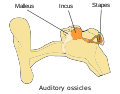 Ossicles
 -
 Head and neck of a human embryo eighteen weeks old, with Meckel's cartilage and hyoid bone exposed.
 -
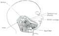
 External and middle ear, opened from the front. Right side.
 -
 Chain of ossicles and their ligaments, seen from the front in a vertical, transverse section of the tympanum.
 -
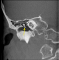 CT image of malleus
 -
 Auditory ossicles. Tympanic cavity. Deep dissection.
 -
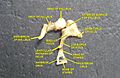
 Auditory ossicles. Incus and malleus. Deep dissection.


See also
- Bone terminology
- Evolution of mammalian auditory ossicles
- Ligaments of malleus
- Terms for anatomical location
References
- ^ OCLC 489078311.
- ^ ISBN 978-0-8089-2306-0.)
{{cite book}}: CS1 maint: multiple names: authors list (link - ^ OCLC 956277358.
- ^ Alidosi, GNP. I dottori Bolognesi di teologia, filosofia, medicina e d'arti liberali dall'anno 1000 per tutto marzo del 1623, Tebaldini, N., Bologna, 1623. http://gallica.bnf.fr/ark:/12148/bpt6k51029z/f35.image#
- ^ Lind, L. R. Studies in pre-Vesalian anatomy. Biography, translations, documents, American Philosophical Society, Philadelphia, 1975. p.40
- ^ Jacopo Berengario da Carpi,Commentaria super anatomia Mundini, Bologna, 1521. https://archive.org/details/ita-bnc-mag-00001056-001
- ^ Niccolo Massa, Liber introductorius anatomiae, Venice, 1536. p.166. https://www.digitale-sammlungen.de/en/view/bsb10151904?page=1
- ^ O'Malley, C.D. Andreas Vesalius of Brussels, 1514-1564. Berkeley: University of California Press, 1964. p. 120
- ISBN 9780688172176.
- OCLC 1196340700.
| National | |
|---|---|
| Other | |