
Mammalian eye
| Eye | |
|---|---|
| Details | |
| Identifiers | |
| Latin | oculus (plural: oculi) |
| Anatomical terminology] | |
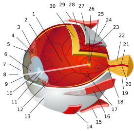
- posterior segment
- ora serrata
- ciliary muscle
- ciliary zonules
- Schlemm's canal
- pupil
- anterior chamber
- cornea
- iris
- lens cortex
- lens nucleus
- ciliary process
- conjunctiva
- inferior oblique muscle
- inferior rectus muscle
- medial rectus muscle
- retinal arteries and veins
- optic disc
- dura mater
- central retinal artery
- central retinal vein
- optic nerve
- vorticose vein
- bulbar sheath
- macula
- fovea
- sclera
- choroid
- superior rectus muscle
- retina
The dimensions of the eyeball vary only 1–2 mm among humans. The vertical axis is 24 mm; the transverse being larger. At birth it is generally 16–17 mm, enlarging to 22.5–23 mm by three years of age. Between then and age 13 the eye attains its mature size. It weighs 7.5 grams and its volume is roughly 6.5 ml. Along a line through the nodal (central) point of the eye is the optic axis, which is slightly five degrees toward the nose from the visual axis (i.e., that going towards the focused point to the fovea).
Three layers
The structure of the
- The fibrous tunic, also known as the tunica fibrosa oculi, is the outer layer of the eyeball consisting of the cornea and sclera.[4] The sclera gives the eye most of its white color. It consists of dense connective tissue filled with the protein collagen to both protect the inner components of the eye and maintain its shape.[5]
- The vascular tunic, also known as the tunica vasculosa oculi or the "uvea", is the middle vascularized layer which includes the iris, ciliary body, and choroid.[4][6][7] The choroid contains blood vessels that supply the retinal cells with necessary oxygen and remove the waste products of respiration. The choroid gives the inner eye a dark color, which prevents disruptive reflections within the eye. The iris is seen rather than the cornea when looking straight in one's eye due to the latter's transparency, the pupil (central aperture of iris) is black because there is no light reflected out of the interior eye. If an ophthalmoscope is used, one can see the fundus, as well as vessels (which supply additional blood flow to the retina) especially those crossing the optic disk—the point where the optic nerve fibers depart from the eyeball—among others[8]
- The nervous tunic, also known as the tunica nervosa oculi, is the inner sensory layer which includes the retina.[4][7]
- Contributing to vision, the retina contains the photosensitive blind". Continuous with the retina are the ciliary epithelium and the posterior epithelium of the iris.
- In addition to the rods and cones, a small proportion (about 1-2% in humans) of the ganglion cells in the retina are themselves photosensitive through the pigment ipRGCshave other functions as well, such as signaling the need for changing the diameter of the pupil in changing light conditions.
- Contributing to vision, the retina contains the photosensitive
Anterior and posterior segments

The mammalian eye can also be divided into two main segments: the
The human eye is not a plain sphere but is like two spheres combined, a smaller, more sharply curved one and a larger lesser curved sphere. The former, the anterior segment is the front sixth
Within the anterior segment are two fluid-filled spaces:
- the anterior chamber between the posterior surface of the cornea (i.e. the corneal endothelium) and the iris.
- the posterior chamber between the iris and the front face of the vitreous.[6]
Some
The posterior segment is the back five-sixths
The radii of the anterior and posterior sections are 8 mm and 12 mm, respectively. The point of junction is called the limbus.
On the other side of the lens is the second humour, the
The tapetum lucidum, in animals that have it, can produce
Some
Extraocular anatomy
Lying over the sclera and the interior of the eyelids is a transparent membrane called the
In many animals, including humans,
In many animals, including humans, eyelashes prevent fine particles from entering the eye. Fine particles can be bacteria, but also simple dust which can cause irritation of the eye, and lead to tears and subsequent blurred vision.[16]
In many species, the eyes are inset in the portion of the skull known as the orbits or eyesockets. This placement of the eyes helps to protect them from injury. For some, the focal fields of the two eyes overlap, providing them with binocular vision. Although most animals have some degree of binocular vision the amount of overlap largely depends on behavioural requirements.
In humans, the eyebrows redirect flowing substances (such as rainwater or sweat) away from the eye.
Function of the mammalian eye
The structure of the mammalian eye owes itself completely to the task of focusing
In the human eye, light enters the pupil and is focused on the retina by the lens. Light-sensitive nerve cells called
Retina
The retina contains three forms of photosensitive cells, two of them important to vision, rods and cones, in addition to the subset of ganglion cells involved in adjusting circadian rhythms and pupil size but probably not involved in vision.
Though structurally and metabolically similar, the functions of rods and cones are quite different. Rod cells are highly sensitive to light, allowing them to respond in dim light and dark conditions; however, they cannot detect color differences. These are the cells that allow humans and other animals to see by moonlight, or with very little available light (as in a dark room). Cone cells, conversely, need high light intensities to respond and have high visual acuity. Different cone cells respond to different wavelengths of light, which allows an organism to see color. The shift from cone vision to rod vision is why the darker conditions become, the less color objects seem to have.
The differences between rods and cones are useful; apart from enabling sight in both dim and light conditions, they have further advantages. The
Rods and cones are both photosensitive, but respond in different ways to different frequencies of light. They contain different pigmented
Differences between the rhodopsin and the iodopsins is the reason why cones and rods enable organisms to see in dark and light conditions — each of the photoreceptor proteins requires a different light intensity to break down into the constituent products. Further,
Furthermore, color is distinguishable due to the different
A small percentage of the ganglion cells in the retina contain
Accommodation

The purpose of the optics of the mammalian eye is to bring a clear image of the visual world onto the retina. Because of limited depth of field of the mammalian eye, an object at one distance from the eye might project a clear image, while an object either closer to or further from the eye will not. To make images clear for objects at different distances from the eye, its optical power needs to be changed. This is accomplished mainly by changing the curvature of the lens. For distant objects, the lens needs to be made flatter; for near objects the lens needs to be made thicker and more rounded.
Water in the eye can alter the optical properties of the eye and blur vision. It can also wash away the tear fluid—along with it the protective lipid layer—and can alter corneal physiology, due to osmotic differences between tear fluid and freshwater. Osmotic effects are made apparent when swimming in freshwater pools, because the osmotic gradient draws water from the pool into the corneal tissue (the pool water is hypotonic), causing edema, and subsequently leaving the swimmer with "cloudy" or "misty" vision for a short period thereafter. The edema can be reversed by irrigating the eye with hypertonic saline which osmotically draws the excess water out of the eye.
References
- ^ "The Eye." Accessed October 23, 2006.
- ^ "General Anatomy of the Eye." Accessed October 23, 2006.
- ^ "Eye Anatomy and Function." Accessed October 23, 2006.
- ^ ISBN 0-7506-9895-0
- ^ X. The Organs of the Senses and the Common Integument. 1c. 1. The Tunics of the Eye. Gray, Henry. 1918. Anatomy of the Human Body
- ^ a b c Cassin, B. and Solomon, S. Dictionary of Eye Terminology. Gainesville, Florida: Triad Publishing Company, 1990.
- ^ a b "Medline Encyclopedia: Eye." Accessed October 25, 2006.
- ^ Encyclopædia Britannica 2006 Ultimate Reference Suite DVD5 Apr. 2008
- PMID 16364902.
Intrinsically photosensitive retinal ganglion cells (ipRGCs) mediate numerous nonvisual phenomena, including entrainment of the circadian clock to light-dark cycles, pupillary light responsiveness, and light-regulated hormone release.
- ^ Ocular Anatomy – Anterior Segment Archived 2008-09-20 at the Wayback Machine
- ^ a b "Departments. Anterior segment." Archived 2006-09-27 at the Wayback Machine Cantabrian Institute of Ophthalmology.
- ^ "Posterior segment anatomy". Archived from the original on 2016-06-03. Retrieved 2008-09-11.
- ^ Vitreoretinal Disease & Surgery – New England Eye Center
- PMID 30137787, retrieved 2022-11-02
- ^ Smith, Michael (2017-08-24). "The eyelid". Vision Eye Institute. Retrieved 2022-11-02.
- ^ Smith, Michael (2017-08-26). "The eyelashes". Vision Eye Institute. Retrieved 2022-11-02.
Vision in animals | ||
|---|---|---|
| Vision | .jpg) | |
| Eyes |
| |
| Evolution | ||
| Coloration | ||
| Related topics | ||