
Mammary gland
| Mammary gland | |
|---|---|
| Details | |
| Precursor | Mesoderm (blood vessels and connective tissue) Ectoderm[3] (cellular elements) |
| Artery | Internal thoracic artery Lateral thoracic artery[1] |
| Vein | Internal thoracic vein Axillary vein[1] |
| Nerve | Supraclavicular nerves Intercostal nerves[2] (lateral and medial branches) |
| Lymph | Pectoral axillary lymph nodes[1] |
| Identifiers | |
| TA98 | A16.0.02.006 |
| TA2 | 7099 |
| FMA | 60088 |
| Anatomical terminology] | |
A mammary gland is an
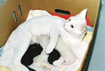
Mammals are divided into 3 groups: prototherians, metatherians, and eutherians. In the case of prototherians, both males and females have functional mammary glands, but their mammary glands are without nipples. These mammary glands are modified sebaceous glands. Concerning metatherians and eutherians, only females have functional mammary glands. Their mammary glands can be termed as breasts or udders. In the case of breasts, each mammary gland has its own nipple (e.g., human mammary glands). In the case of udders, pairs of mammary glands comprise a single mass, with more than one nipple (or teat) hanging from it. For instance, cows and buffalo udders have two pairs of mammary glands and four teats, whereas sheep and goat udders have one pair of mammary glands with two teats protruding from the udder. Each gland produces milk for a single teat. These mammary glands are modified sweat glands.
Structure
The basic components of a mature mammary gland are the
All the milk-secreting tissue leading to a single lactiferous duct is collectively called a "simple mammary gland"; in a "complex mammary gland", all the simple mammary glands serve one nipple. Humans normally have two complex mammary glands, one in each breast, and each complex mammary gland consists of 10–20 simple glands. The opening of each simple gland on the surface of the nipple is called a "pore."
Maintaining the correct polarized morphology of the lactiferous duct tree requires another essential component – mammary epithelial cells extracellular matrix (ECM) which, together with adipocytes, fibroblast, inflammatory cells, and others, constitute mammary stroma.[6] Mammary epithelial ECM mainly contains myoepithelial basement membrane and the connective tissue. They not only help to support mammary basic structure, but also serve as a communicating bridge between mammary epithelia and their local and global environment throughout this organ's development.[7][8]
Histology


A mammary gland is a specific type of apocrine gland specialized for manufacture of colostrum when giving birth. Mammary glands can be identified as apocrine because they exhibit striking "decapitation" secretion. Many sources assert that mammary glands are modified sweat glands.[9][10][11] Some authors dispute that and argue instead that they are sebaceous glands.[9]
Development
Mammary glands develop during different growth cycles. They exist in both sexes during the embryonic stage, forming only a rudimentary duct tree at birth. In this stage, mammary gland development depends on systemic (and maternal) hormones,
Biochemistry
During embryonic development, IGF-1 levels are low, and gradually increase from birth to puberty.
Androgens such as testosterone inhibit estrogen-mediated mammary gland development (e.g., by reducing local ER expression) through activation of androgen receptors expressed in mammary gland tissue,[29][30] and in conjunction with relatively low estrogen levels, are the cause of the lack of developed mammary glands in males.[31]
Timeline
Before birth
Mammary gland development is characterized by the unique process by which the
Developmentally, mammary gland epithelium is constantly produced and maintained by rare epithelial cells, dubbed as mammary progenitors which are ultimately thought to be derived from tissue-resident stem cells.[33]
The primitive (stem) cells are detected in embryo and their numbers increase steadily during development[35]
Growth
By the pubertal stage, the mammary ducts have invaded to the end of the mammary fat pad. At this point, the terminal end buds become less proliferative and decrease in size. Side branches form from the primary ducts and begin to fill the mammary fat pad. Ductal development decreases with the arrival of sexual maturity and undergoes estrous cycles (proestrus, estrus, metestrus, and diestrus). As a result of estrous cycling, the mammary gland undergoes dynamic changes where cells proliferate and then regress in an ordered fashion.[36]
Pregnancy
During
Postmenopausal
During
Physiology
Hormonal control
Lactiferous duct development occurs in females in response to circulating
There is preliminary evidence that soybean intake mildly stimulates the breast glands in pre- and postmenopausal women.[44]
Pregnancy
Secretory alveoli develop mainly in pregnancy, when rising levels of
Weaning
During weaning, decreased prolactin, missing mechanical stimulation (baby suckling), and changes in osmotic balance caused by milk stasis and leaking of tight junctions cause cessation of milk production. It is the (passive) process of a child or animal ceasing to be dependent on the mother for nourishment. In some species there is complete or partial
Clinical significance
Other mammals
General
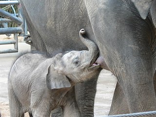
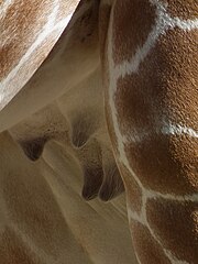
The breasts of female humans vary from most other mammals that tend to have less conspicuous mammary glands. The number and positioning of mammary glands varies widely in different mammals. The protruding teats and accompanying glands can be located anywhere along the two
| Species[57] | Anterior ( thoracic )
|
Intermediate ( abdominal )
|
Posterior (inguinal) |
Total |
|---|---|---|---|---|
| Goat, sheep, horse guinea pig |
0 | 0 | 2 | 2 |
| Cattle | 0 | 0 | 4 | 4 |
| Cat | 2 | 2 | 4 | 8 |
| Dog[58] | 4 | 2 | 2 or 4 | 8 or 10 |
| Mouse | 6 | 0 | 4 | 10 |
| Rat | 6 | 2 | 4 | 12 |
| Pig | 6 | 6 | 6 | 18 |
primates
|
2 | 0 | 0 | 2 |
| Virginia opossum[55][56] | 0 | 0 | 13 | 13 |
| Southern red-sided opossum[59] | 0 | 0 | 25 to 27 | 25 to 27 |
Male mammals typically have rudimentary mammary glands and nipples, with a few exceptions: male mice do not have nipples,[60] male marsupials do not have mammary glands,[61] and male horses lack nipples.[62] The male dayak fruit bat has lactating mammary glands.[63] Male lactation occurs infrequently in some species.[64]
Mammary glands are true
Evolution
There are many theories on how mammary glands evolved. For example, it is thought that the mammary gland is a transformed sweat gland, more closely related to
Lactation is thought to have developed long before the evolution of the mammary gland and mammals; see evolution of lactation.
Additional images
-
 Cross section of the breast of a human female
 -

-

-
-

-

-
-
 Dromedary camel
 -


See also
List of distinct cell types in the adult human body
References
- ^ .
- ISBN 9781437735901.
- ^ Gray, Henry (1918). Anatomy of the Human Body.
- PMID 18101061.
- PMID 26855906.
- ^ S2CID 9089976.
- ^ PMID 12004111.
- PMID 20649458.
- ^ a b Ackerman (2005) ch.1 Apocrine Units Archived 21 April 2011 at the Wayback Machine
- ^ Moore (2010) ch.1 Thorax, p. 99
- ISBN 9783540536666.
- PMID 9477327.
- PMID 16168142.
- PMID 27544910.
- ^ PMID 20739412.
- ^ S2CID 30440069.
- ^ S2CID 41667675.
- ^ PMID 10537134.
- ^ S2CID 25656770.
- S2CID 24786346.
- PMID 16322320.
- PMID 17659070.
- PMID 21934211.
- ISBN 978-0-7020-3489-3.
- PMID 20554705.
- ISBN 978-1-4511-4870-1.
- ^ PMID 17595785.
- ISBN 978-0-12-387584-6.
- ^ PMID 9098173.
- S2CID 17172449.
- PMID 24872741.
- PMID 6053715.
- PMID 31267556.
- PMID 16168142.
- PMID 23624881.
- S2CID 36670489.
- PMID 16524451.
- PMID 16916375.
- PMID 14680479.
- PMID 12975354.
- PMID 10662785.
- PMID 9472007.
- PMID 12460932.
- PMID 8896889.
- S2CID 2319046.
- PMID 10733525.
- ^ PMID 1955479.
- ^ PMID 7730398.
- PMID 1168727.
- PMID 19261986.
- S2CID 5926448.
- PMID 17228094.
- PMID 11801722.
- PMID 18442412.

- ^ a b "With the Wild Things – Transcripts". Digitalcollections.fiu.edu. Archived from the original on 23 March 2013. Retrieved 5 April 2013.
- ^ a b Stockard, Mary (2005) Raising Orphaned Baby Opossums. Alabama Wildlife Center.
- ISBN 978-0-13-046256-5.
- ^ Dog breeds vary in the number of mammary glands: larger breeds tend to have 5 pairs, smaller breeds have 4 pairs.[citation needed]
- ^ P Smith 2008 Red-Sided Short-Tailed Opossum. Fauna Paraguay
- PMID 18832580.
- ISBN 978-1-139-45742-2.
- PMID 33280071.
- S2CID 4369716.
- PMID 19100649.
- PMID 19549750.
- ^ "BBC News – The goats with spider genes and silk in their milk". bbc.co.uk. 17 January 2012. Retrieved 26 April 2012.
- S2CID 8319185.
- ^ Lactating on Eggs. Smithsonian National Zoo, 14 July 2003.
- S2CID 25806501.
- ^ Breast beginnings. scienceblogs.com
- PMID 16700061.
- PMID 20565255.
- S2CID 84089647.
Bibliography
- Ackerman, A. Bernard; Almut Böer; Bruce Bennin; Geoffrey J. Gottlieb (2005). Histologic Diagnosis of Inflammatory Skin Diseases An Algorithmic Method Based on Pattern Analysis. Ardor Scribendi. ISBN 978-1-893357-25-9. Archived from the originalon 21 April 2011.
- Moore, Keith L. et al. (2010) Clinically Oriented Anatomy 6th Ed
External links
- Comparative Mammary Gland Anatomy by W. L. Hurley
- On the anatomy of the breast by Sir Astley Paston Cooper (1840). Numerous drawings, in the public domain.