
Medial pterygoid muscle
| Medial pterygoid | |
|---|---|
elevates mandible, closes jaw, helps lateral pterygoids in moving the jaw from side to side | |
| Identifiers | |
| Latin | musculus pterygoideus medialis, musculus pterygoideus internus |
| TA98 | A04.1.04.009 |
| TA2 | 2113 |
| FMA | 49011 |
| Anatomical terms of muscle] | |
The medial pterygoid muscle (or internal pterygoid muscle) is a thick, quadrilateral muscle of the
Structure
The medial pterygoid muscle consists of two heads. The bulk of the muscle arises as a deep head from just above the medial surface of the
Its fibers pass downward, lateral, and posterior, and are inserted, by a strong tendinous lamina, into the lower and back part of the medial surface of the
Nerve supply
The medial pterygoid muscle is supplied by the medial pterygoid nerve, a branch of the mandibular nerve, itself a branch of the trigeminal nerve (V). This also supplies the tensor tympani muscle and the tensor veli palatini muscle. The medial pterygoid nerve is a main trunk from the mandibular nerve, before the division of the trigeminal nerve - this is unlike the lateral pterygoid muscle, and all other muscles of mastication which are supplied by the anterior division of the mandibular nerve.
Function
The medial pterygoid muscle has functions including elevating the mandible (closing the mouth), protruding the mandible, mastication (especially for when the maxillary teeth and the mandibular teeth are close together),[1] and excursing the mandible (contralateral excursion occurs with unilateral contraction).
Additional images
-
 Position of medial pterygoid muscle (red).
 -
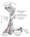 Left palatine bone. Posterior aspect. Enlarged.
 -
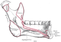 Mandible. Inner surface. Side view.
 -
 Plan of branches of internal maxillary artery.
 -
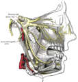 Distribution of the maxillary and mandibular nerves, and the submaxillary ganglion.
 -
 Mandibular division of trifacial nerve, seen from the middle line.
 -
 Muscles of the pharynx, viewed from behind, together with the associated vessels and nerves.
 -
 Deep dissection. Anterior view.


-
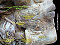
 Medial pterygoid muscle
 -
 Medial pterygoid muscle
 -
 Medial pterygoid muscle
 -
 Medial pterygoid muscle
 -
 Medial pterygoid muscle
 -
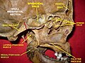
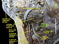
 Infratemporal fossa. Lingual and inferior alveolar nerve. Deep dissection. Anterolateral view


References
![]() This article incorporates text in the public domain from page 387 of the 20th edition of Gray's Anatomy (1918)
This article incorporates text in the public domain from page 387 of the 20th edition of Gray's Anatomy (1918)
- PMID 3458914.
External links
- MedicalMnemonics.com: 70
- "Anatomy diagram: 25420.000-1". Roche Lexicon - illustrated navigator. Elsevier. Archived from the original on 2015-02-26.