Mediastinum
This article needs additional citations for verification. (September 2014) |
| Mediastinum | |
|---|---|
high resolution computed tomography of the thorax, with mediastinum marked in blue. | |
 Mediastinum. The division between superior and inferior is at the sternal angle. | |
| Details | |
| Identifiers | |
| Latin | mediastinum[1] |
| MeSH | D008482 |
| TA98 | A07.1.02.101 |
| TA2 | 3333 |
| FMA | 9826 |
| Anatomical terminology] | |
The mediastinum (from
Anatomy
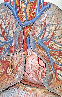
The mediastinum lies within the

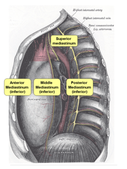
The mediastinum can be divided into an upper (or superior) and lower (or inferior) part:
- The superior mediastinum starts at the superior thoracic aperture and ends at the thoracic plane.
- The inferior mediastinum from this level to the diaphragm. This lower part is subdivided into three regions, all relative to the pericardium – the anterior mediastinum being in front of the pericardium, the middle mediastinum contains the pericardium and its contents, and the posterior mediastinum being behind the pericardium.[6]
Thoracic plane
The transverse thoracic plane, thoracic plane, plane of Louis or plane of Ludwig is an important anatomical plane at the level of the sternal angle and the T4/T5 intervertebral disc.[8][9][10] It serves as an imaginary boundary that separates the superior and inferior mediastinum.[8][9][10]
A number of important anatomical structures and transitions occur at the level of the thoracic plane, including:
- The bronchi.
- The pulmonary trunkbelow.
- The starting of the cardiac plexus.
- The azygos vein arching over the right main bronchus and joining into the superior vena cava.
- The thoracic duct crossing the midline from right to left behind the esophagus
- The end of the pretracheal and prevertebral fasciae.
Superior mediastinum
The superior mediastinum is bounded:
- superiorly by the thoracic inlet, the upper opening of the thorax;
- inferiorly by the transverse thoracic plane. which is an imaginary plane passing from the sternal angle anteriorly to the lower border of the body of the 4th thoracic vertebra posteriorly;
- laterally by the pleurae;
- anteriorly by the manubriumof the sternum;
- posteriorly by the first four thoracic vertebral bodies.
Contents
|
|---|
 
|
Inferior mediastinum
Anterior inferior mediastinum
Is bounded:
- laterally by the pleurae;
- posteriorly by the pericardium;[4]
- anteriorly by the costal cartilages.
Contents
|
|---|
|
Middle inferior mediastinum
Bounded:
Contents
|
|---|
|
Posterior inferior mediastinum
Is bounded:
- Anteriorly by (from above downwards): bifurcation of trachea; pulmonary vessels; fibrous pericardium and posterior sloping surface of diaphragm
- Inferiorly by the thoracic surface of the diaphragm(below);
- Superiorly by the transverse thoracic plane;
- Posteriorly by the bodies of the thoracic vertebra (behind);[4]
- Laterally by the mediastinal pleura(on either side).
Contents
|
|---|
|
-
A transverse section of theposterior mediastinum.
Clinical significance

The mediastinum is frequently the site of involvement of various
- Anterior mediastinum: substernal .
- Middle mediastinum: lymphadenopathy, metastatic disease such as from small cell carcinoma from the lung.
- Posterior mediastinum: Neurogenic tumors, either from the nerve sheath (mostly benign) or elsewhere (mostly malignant).
Mediastinitis is inflammation of the tissues in the mediastinum, usually bacterial and due to rupture of organs in the mediastinum. As the infection can progress very quickly, this is a serious condition.
Widening
| Widened mediastinum | |
|---|---|
| Other names | Mediastinal widening |
achalasia | |
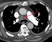
Widened mediastinum/mediastinal widening is where the mediastinum has a width greater than 6 cm on an upright PA
A widened mediastinum can be indicative of several pathologies:[12][13]
- aortic aneurysm[14]
- aortic dissection[15]
- aortic unfolding
- aortic rupture
- hilar lymphadenopathy
- anthrax inhalation - a widened mediastinum was found in 7 of the first 10 victims infected by anthrax (Bacillus anthracis) in 2001.[16]
- esophageal rupture - presents usually with pneumomediastinum and pleural effusion. It is diagnosed with water-soluble swallowed contrast.
- mediastinal mass
- mediastinitis
- cardiac tamponade[17]
- pericardial effusion
- thoracic vertebrae fracturesin trauma patients.
See also
- Mediastinum testis (unrelated structure in the scrotum)
- Mediastinal germ cell tumor
- Mediastinitis
- Mediastinal tumor
- List of anatomy mnemonics#Mediastinum
References
![]() This article incorporates text in the public domain from page 1090 of the 20th edition of Gray's Anatomy (1918)
This article incorporates text in the public domain from page 1090 of the 20th edition of Gray's Anatomy (1918)
- ^ "A index W LA".
- ^ "Mediastinum dictionary definition - mediastinum defined". www.yourdictionary.com.
- ISBN 978-0-12-370879-3, retrieved 2020-11-17
- ^ ISBN 978-1-4160-4208-2, retrieved 2020-11-17
- ISBN 978-1-4557-3383-5, retrieved 2020-11-17
- ISBN 978-0-7020-3425-1, retrieved 2020-11-17
- ^ Goodman, Lawrence. Felson's Principles of Chest Roentgenology.
- ^ a b "Thoracic Wall, Pleura, and Pericardium – Dissector Answers". Archived from the original on September 1, 2012.
- ^ a b "Cell Biology and Anatomy - School of Medicine - University of South Carolina". dba.med.sc.edu. Archived from the original on 2006-09-05. Retrieved 2014-09-22.
- ^ a b "UAMS Department of Neurobiology and Developmental Sciences - Topographical Anatomy of the Thorax". anatomy.uams.edu. Archived from the original on 2004-08-17. Retrieved 2014-09-22.
- ^ D'Souza, Donna. "Thoracic aortic injury | Radiology Reference Article | Radiopaedia.org". radiopaedia.org.
- PMID 16034263.
- PMID 2357135.
- PMID 1113190.
- PMID 14715319.
- PMID 11747719.
- ISBN 978-1-56053-506-5. Retrieved 19 April 2010.
External links
- Anatomy figure: 21:01-03 at Human Anatomy Online, SUNY Downstate Medical Center – "Divisions of the mediastinum."
- Anatomy figure: 21:02-03 at Human Anatomy Online, SUNY Downstate Medical Center – "The anatomical divisions of the inferior mediastinum."
- thoraxlesson3 at The Anatomy Lesson by Wesley Norman (Georgetown University) – "Subdivisions of the Thoracic Cavity"

