
Mental foramen
| Mental foramen | |
|---|---|
Mandible. Outer surface. Side view. (Mental foramen visible at left.) | |
| Details | |
| Part of | mandible |
| Identifiers | |
| Latin | foramen mentale |
| MeSH | D000080383 |
| TA98 | A02.1.15.007 |
| TA2 | 845 |
| FMA | 53171 |
| Anatomical terms of bone] | |

The mental foramen is one of two
Structure
The mental foramen is located on the anterior surface of the
Variation
The mental foramen descends slightly in
The mental foramen is in line with the longitudinal axis of the 2nd premolar in 63% of people.[3] It generally lies at the level of the vestibular fornix and about a finger's breadth above the inferior border of the mandible.
In the general population, 17% of mandibles have an additional mental foramen or foramina on at least one side,[3] while 4% of the mandibles show multiple mental foramina on both sides. Most are unequal in size, often with a single large foramen while any others are smaller.[3] An incisive mental foramen is observed in 1% of the side of the mandible.[3]
Clinical significance
The mental nerve may be anaesthetized as it leaves the mental foramen.[1] This causes loss of sensation to the lower lip and chin on the same side.[1]
Additional images
-
 Side view of the skull.
 -
 The skull from the front.
 -
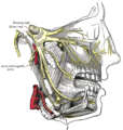 Distribution of the maxillary and mandibular nerves, and the submaxillary ganglion.
 -
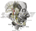 Mandibular division of the trifacial nerve.
 -
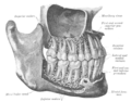 The permanent teeth, viewed from the right.


See also
References
- ^ ISBN 978-0-7020-2980-6.
- PMID 8558356.
- ^ a b c d Jaffar, A. A.; Al-Zubaidi, A. F.; Al-Salihi, A. R. (2002). "Anatomical features of clinical significance in dry mandibles". Iraqi Dental Journal. 29: 99–118. Archived from the original on 2008-12-29.
External links
- cranialnerves at The Anatomy Lesson by Wesley Norman (Georgetown University) (V)
- Diagram at uni-mainz.de
This human musculoskeletal system article is a stub. You can help Wikipedia by expanding it. |