
Metatarsal bones
(Redirected from
Metatarsal
)| Metatarsal bones | |
|---|---|
 Skeleton of foot. Superior view. Metatarsals shown in green | |
 Skeleton of left foot. Lateral aspect. Metatarsals shown in purple | |
| Details | |
| Identifiers | |
| Latin | metatarsus pl. ossa metatarsi (also: ossa metatarsalia) |
| MeSH | D008682 |
| TA98 | A02.5.17.001 |
| TA2 | 1495 |
| FMA | 71340 |
| Anatomical terms of bone | |
The metatarsal bones or metatarsus (pl.: metatarsi) are a group of five
metacarpal bones of the hand. The lengths of the metatarsal bones in humans are, in descending order, second, third, fourth, fifth, and first.[1] A bovine hind leg has two metatarsals.[2]
Structure
The five metatarsals are
plantar surface is grooved antero-posteriorly for the passage of the flexor tendons, and marked on either side by an articular eminence continuous with the terminal articular surface.[4]
During growth, the growth plates are located distally on the metatarsals, except on the first metatarsal where it is located proximally. Yet it is quite common to have an accessory growth plate on the distal first metatarsal.[5]
Articulations

The base of each metatarsal bone articulates with one or more of the tarsal bones at the
intermetatarsal joints
- The intermediate cuneiform.[6]
- the cuneiforms.[6]
- the lateral cuneiform.[6]
- the fourth with the lateral cuneiform and the cuboid.[6]
- The fifth with the cuboid.[6]
Muscle attachments
 |
 |
| Muscle | Direction | Attachment[7] |
| Tibialis anterior | Insertion | Basis of first metatarsal |
Peroneous tertius |
Insertion | Dorsal side basis of fifth metatarsal |
Peroneous longus |
Insertion | Tuberosity of first metatarsal |
Peroneous brevis |
Insertion | Tuberosity of fifth metatarsal |
| Horizontal head of adductor hallucis | Origin | Deep transverse metatarsal ligament |
Flexor digiti minimi brevis |
Origin | Basis of fifth metatarsal |
| Plantar interossei | Origin | Medial side of third, fourth and fifth metatarsal |
| Dorsal interossei | Origin | First to fifth metatarsal |
Clinical significance
Injuries

The metatarsal bones are often broken by
oversupinated during locomotion.[9]
Protection from injuries can be given by the use of safety footwear which can use built-in or removable metatarsal guards.
Additional images
-
 X-ray of foot.
 -
 Skeleton of left foot. Medial aspect.
 -
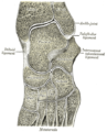 Oblique section of left intertarsal and tarsometatarsal articulations, showing the synovial cavities.
 -
 Ankle and tarsometarsal joints, showing bones of foot. Deep dissection.
 -
 Safety footwear with removable metatarsal guard.


See also
- Arches of the foot
- Ball (foot)
- Bone terminology
- Terms for anatomical location
- Jones fracture
- Lisfranc injury
- Morton's toe
Notes
- ISBN 978-87-628-0307-7.
- ^ "Identification – cattle hock bone |".
- ^ Platzer 2004, p. 220
- ^ Gray's 1918, 6d. 2. The Metatarsus
- PMID 2681682.
- ^ a b c d e Platzer 2004, p. 218
- ISBN 978-87-628-0307-7.
- ^ Bill, Mills (11 December 2010). "Sock boffs may have cured metatarsal woes for Rooney and Co". www.mirrorfootball.co.uk. Retrieved 12 December 2010.
- ^ Perron, Andrew D. (2005-11-23). "Metatarsal Stress Fracture". Retrieved 2007-09-13.
References
- Platzer, Werner (2004). Color Atlas of Human Anatomy, Vol. 1: Locomotor System (5th ed.). Thieme. ISBN 3-13-533305-1.
- Gray, Henry (1918). "6d. 2. The Metatarsus". Anatomy of the Human Body. Bartleby.com.
External links
Wikimedia Commons has media related to Metatarsus.
- Anatomy figure: 16:01-05 at Human Anatomy Online, SUNY Downstate Medical Center
| National | |
|---|---|
| Other | |