
Mitral valve
| Mitral valve | |
|---|---|
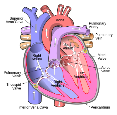 Anterior (frontal) view of the opened heart. White arrows indicate normal blood flow. (Mitral valve labeled at center right.) | |
 Base of ventricles exposed by removal of the atria. (Bicuspid (mitral) valve visible at bottom left.
Tricuspid valve visible at bottom right.) | |
| Details | |
| Identifiers | |
| Latin | valva atrioventricularis sinistra, valva mitralis, valvula bicuspidalis |
| MeSH | D008943 |
| TA98 | A12.1.04.003 |
| TA2 | 3987 |
| FMA | 7235 |
| Anatomical terminology | |
The mitral valve (
In normal conditions, blood flows through an open mitral valve during diastole with contraction of the left atrium, and the mitral valve closes during systole with contraction of the left ventricle. The valve opens and closes because of pressure differences, opening when there is greater pressure in the left atrium than ventricle and closing when there is greater pressure in the left ventricle than atrium.[2]
In abnormal conditions, blood may flow backward through the valve (
Structure


The mitral valve is typically 4 to 6 square centimetres (0.62 to 0.93 sq in) in area and sits in the left heart between the left atrium and the left ventricle.[5] It has two cusps: an anterior one, and a posterior one.[6] The opening of the mitral valve is surrounded by a fibrous ring known as the mitral annulus.[citation needed] The anterior cusp attaches to one third of the circumverence of the annulus, and the posterior cusp attaches to the remaining two thirds of its circumference. Occasionally, the anterior and posterior cusps close the orifice incompletely and a small additional accessory cusp is present to fill the interval. The anterior cusp is thicker and more rigid than the posterior one,[6] and covers approximately two-thirds of the valve.[citation needed] The anterior cusp intervenes between the mitral and aortic orifices.[6] Although the anterior leaflet takes up a larger part of the ring and rises higher, the posterior leaflet has a larger surface area.[citation needed]
Leaflets

In Carpentier's classification of a mitral valve, both the posterior and anterior mitral valve leaflets are divided into eight segments: P3 (medial scallop), P2 (middle scallop), P1 (lateral scallop), A3 (anteromedial segment), A2 (anteromedial), A1 (anterolateral), PMC (posteromedial commissure), ALC (anterolateral commissure).[7] Mitral leaflet thickness is usually about 1 mm but sometimes can range from 3–5 mm.[7][8]
Chordae tendineae


The valve leaflets are prevented from prolapsing into the left atrium by the action of chordae tendineae. The chordae tendineae are inelastic tendons attached at one end to papillary muscles in the left ventricle, and at the other to the valve cusps. Papillary muscles are finger-like projections from the wall of the left ventricle.
When the left ventricle contracts, the pressure in the ventricle forces the valve to close, while the tendons keep the leaflets coapting together and prevent the valve from opening in the wrong direction (thus preventing blood flowing back to the left atrium). Each chord has a different thickness. The thinnest ones are attached to the free leaflet margin, whereas the thickest ones (strut chords) are attached further from the free margin. This disposition has important effects on systolic stress distribution physiology.[9]
Annulus
The mitral annulus is a
The normal diameter of the mitral annulus is 2.7 to 3.5 centimetres (1.1 to 1.4 in), and the circumference is 8 to 9 centimetres (3.1 to 3.5 in). Microscopically, there is no evidence of an annular structure anteriorly, where the mitral valve leaflet is contiguous with the posterior aortic root.[12]
Function
During
After the E wave, there is a period of slow filling of the ventricle.
Left atrial contraction (

The mitral annulus changes in shape and size during the cardiac cycle. It is smaller at the end of atrial systole due to the contraction of the left atrium around it, like a
Clinical significance
Disease

There are some valvular heart diseases that affect the mitral valve. Mitral stenosis is a narrowing of the valve. This can be heard as an opening snap; a heart sound which is not normally present.
Classic mitral valve prolapse is caused by an excess of connective tissue that thickens the spongiosa layer of the cusp and separates collagen bundles in the fibrosa. This weakens the cusps and adjacent tissue, resulting in an increased cuspal area and lengthening of the chordae tendineae. Elongation of the chordae tendineae often causes rupture, commonly to the chordae attached to the posterior cusp. Advanced lesions—also commonly involving the posterior leaflet—lead to leaflet folding, inversion, and displacement toward the left atrium.[14]
A valve prolapse can result in
There are also some rarer forms of congenital mitral valve disease that are often associated with other congenital heart anomalies. Parachute mitral valve occurs when all chordae tendineae of the mitral valve are abnormally attached to a single (or fused) papillary muscle. Straddling Mitral Valve occurs when the mitral valve's chordal attachments straddles, or goes through, a ventricular septal defect (VSD) and so has chordae originating on both sides of the ventricular septum. Mitral valve agenesis is very rare, defined as an absence or minimal presence of both mitral valve leaflets (complete agenesis) or one of the leaflets (partial agenesis).[15]
Surgery can be performed to
Rarely there can be a severe form of calcification of the mitral valve annulus that can be mistaken for an intracardiac mass or thrombus.[16]
Mitral disease can be classified using Carpentier's classification which is based on the leaflet motion. Type I pertains to normal leaflet motion. Whereas, disease of the valve is categorized to primary mitral regurgitation or secondary mitral regurgitation based on the regurgitant etiology. Type II pertains to excessive leaflet motion leading to leaflet prolapse. Common causes include, but is not limited to, Barlow disease, myxomatous degeneration, inflammation, and papillary muscle rupture. Type III pertains to restrictive motion of the leaflets. Type IIIa pertains to restrictive motion during systole and diastole. Type IIIb pertains to restrictive motion during systole.[17]
Investigation
The closing of the mitral valve and the tricuspid valve constitutes the
The mitral valve is often also investigated using an
Etymology
The word mitral comes from
, meaning "point", reflecting the dual-flap shape of the valve.Gallery
-
 The human heart, viewed from the front. The mitral valve is visible on the right as the "bicuspid valve"
 -
-
 Mitral valve, viewed in a cadaver specimen from within the left atrium.


See also
- Heart
- Heart rhythm
- Electrocardiography
References
- )
- )
- OCLC 1054224262.
- )
- S2CID 240154628.
- ^ ISBN 978-0-7295-3752-0.
- ^ PMID 23960615.
- PMID 20435783.
- PMID 10901521.
- PMID 23962462.
- PMID 5046018.
- ISBN 9781416058922.
- PMID 12578332. Archived from the originalon 2011-07-13. Retrieved 2010-03-04.
- PMID 12439384. Archived from the originalon 2014-09-03. Retrieved 2015-01-01.
- PMID 21944149.
- PMID 24791181.
- ^ Patrizio Lancellotti, Philippe Pibarot, John Chambers, Giovanni La Canna, Mauro Pepi, Raluca Dulgheru, Mark Dweck, Victoria Delgado, Madalina Garbi, Mani A Vannan, David Montaigne, Luigi Badano, Pal Maurovich-Horvat, Gianluca Pontone, Alec Vahanian, Erwan Donal, Bernard Cosyns, the Scientific Document Committee of the European Association of Cardiovascular Imaging, Multi-modality imaging assessment of native valvular regurgitation: an EACVI and ESC council of valvular heart disease position paper, European Heart Journal - Cardiovascular Imaging, Volume 23, Issue 5, May 2022, Pages e171–e232, https://doi.org/10.1093/ehjci/jeab253
Further reading
- Ingels, Neil B.; Karlsson, Matts (21 January 2016). Mitral Valve Mechanics (PDF). Linköping University Electronic Press. ISBN 9789176859520. Archived(PDF) from the original on 19 May 2022.
External links
- Anatomy figure: 20:07-03 at Human Anatomy Online, SUNY Downstate Medical Center—"Valves of the heart"
- Cardiac Valve Animations—Perioperative Interactive Education Group