
Molecular models of DNA
Molecular models of DNA structures are representations of the
The more advanced, computer-based
History

From the very early stages of structural studies of DNA by
Structural information is generated from X-ray diffraction studies of oriented DNA fibers with the help of molecular models of DNA that are combined with crystallographic and mathematical analysis of the X-ray patterns.
The first reports of a double helix molecular model of B-DNA structure were made by James Watson and Francis Crick in 1953.[5][6] That same year, Maurice F. Wilkins, A. Stokes and H.R. Wilson, reported the first X-ray patterns of in vivo B-DNA in partially oriented salmon sperm heads.[7]
The development of the first correct double helix molecular model of DNA by Crick and Watson may not have been possible without the biochemical evidence for the nucleotide base-pairing ([A---T]; [C---G]), or Chargaff's rules.[8][9][10][11][12][13] Although such initial studies of DNA structures with the help of molecular models were essentially static, their consequences for explaining the in vivo functions of DNA were significant in the areas of protein biosynthesis and the quasi-universality of the genetic code. Epigenetic transformation studies of DNA in vivo were however much slower to develop despite their importance for embryology, morphogenesis and cancer research. Such chemical dynamics and biochemical reactions of DNA are much more complex than the molecular dynamics of DNA physical interactions with water, ions and proteins/enzymes in living cells.
Importance
An old standing dynamic problem is how DNA "self-replication" takes place in living cells that should involve transient uncoiling of supercoiled DNA fibers. Although DNA consists of relatively rigid, very large elongated biopolymer molecules called fibers or chains (that are made of repeating
Such varying molecular geometries can also be computed, at least in principle, by employing ab initio quantum chemistry methods that can attain high accuracy for small molecules, although claims that acceptable accuracy can be also achieved for polynuclelotides, and DNA conformations, were recently made on the basis of vibrational circular dichroism (VCD) spectral data. Such quantum geometries define an important class of ab initio molecular models of DNA which exploration has barely started, especially related to results obtained by VCD in solutions. More detailed comparisons with such ab initio quantum computations are in principle obtainable through 2D-FT NMR spectroscopy and relaxation studies of polynucleotide solutions or specifically labeled DNA, as for example with deuterium labels.
In an interesting twist of roles, the DNA molecule was proposed to be used for quantum computing via DNA. Both DNA nanostructures and DNA computing biochips have been built.
Fundamental concepts


The chemical structure of DNA is insufficient to understand the complexity of the 3D structures of DNA. In contrast, animated molecular models allow one to visually explore the three-dimensional (3D) structure of DNA. The DNA model shown (far right) is a space-filling, or CPK, model of the DNA double helix. Animated molecular models, such as the wire, or skeletal, type shown at the top of this article, allow one to visually explore the three-dimensional (3D) structure of DNA. Another type of DNA model is the space-filling, or CPK, model.
The hydrogen bonding dynamics and proton exchange is very different by many orders of magnitude between the two systems of fully hydrated DNA and water molecules in ice. Thus, the DNA dynamics is complex, involving nanosecond and several tens of picosecond time scales, whereas that of liquid ice is on the picosecond time scale, and that of proton exchange in ice is on the millisecond time scale. The proton exchange rates in DNA and attached proteins may vary from picosecond to nanosecond, minutes or years, depending on the exact locations of the exchanged protons in the large biopolymers.
A simple harmonic oscillator 'vibration' is only an oversimplified dynamic representation of the longitudinal vibrations of the DNA intertwined helices which were found to be anharmonic rather than harmonic as often assumed in quantum dynamic simulations of DNA.
DNA structure
The structure of DNA shows a variety of forms, both double-stranded and single-stranded. The mechanical properties of DNA, which are directly related to its structure, are a significant problem for cells. Every process which binds or reads DNA is able to use or modify the mechanical properties of DNA for purposes of recognition, packaging and modification. The extreme length (a chromosome may contain a 10 cm long DNA strand), relative rigidity and helical structure of DNA has led to the evolution of histones and of enzymes such as topoisomerases and helicases to manage a cell's DNA. The properties of DNA are closely related to its molecular structure and sequence, particularly the weakness of the hydrogen bonds and electronic interactions that hold strands of DNA together compared to the strength of the bonds within each strand.
Experimental methods which can directly measure the mechanical properties of DNA are relatively new, and high-resolution visualization in solution is often difficult. Nevertheless, scientists have uncovered large amount of data on the mechanical properties of this polymer, and the implications of DNA's mechanical properties on cellular processes is a topic of active current research.
The DNA found in many cells can be macroscopic in length: a few centimetres long for each human chromosome. Consequently, cells must compact or package DNA to carry it within them. In eukaryotes this is carried by spool-like proteins named histones, around which DNA winds. It is the further compaction of this DNA-protein complex which produces the well known mitotic eukaryotic chromosomes.
In the late 1970s, alternate
DNA structure determination using molecular modeling and DNA X-ray patterns


After DNA has been separated and purified by standard biochemical methods, one has a sample in a jar much like in the figure at the top of this article. Below are the main steps involved in generating structural information from X-ray diffraction studies of oriented DNA fibers that are drawn from the hydrated DNA sample with the help of molecular models of DNA that are combined with crystallographic and mathematical analysis of the X-ray patterns.
Paracrystalline lattice models of B-DNA structures
A
Liquid crystals also have paracrystalline rather than crystalline structures.
Highly hydrated B-DNA occurs naturally in living cells in such a paracrystalline state, which is a dynamic one despite the relatively rigid DNA double helix stabilized by parallel hydrogen bonds between the nucleotide base-pairs in the two complementary, helical DNA chains (see figures). For simplicity most DNA molecular models omit both water and ions dynamically bound to B-DNA, and are thus less useful for understanding the dynamic behaviors of B-DNA in vivo. The physical and mathematical analysis of X-ray[16][17] and spectroscopic data for paracrystalline B-DNA is thus far more complex than that of crystalline, A-DNA X-ray diffraction patterns. The paracrystal model is also important for DNA technological applications such as DNA nanotechnology. Novel methods that combine X-ray diffraction of DNA with X-ray microscopy in hydrated living cells are now also being developed.[18]
Genomic and biotechnology applications of DNA molecular modeling
There are various uses of DNA molecular modeling in Genomics and Biotechnology research applications, from DNA repair to PCR and DNA nanostructures. Two-dimensional DNA junction arrays have been visualized by Atomic force microscopy.[19]
DNA molecular modeling has various uses in genomics and biotechnology, with research applications ranging from DNA repair to PCR and DNA nanostructures. These include computer molecular models of molecules as varied as RNA polymerase, an E. coli, bacterial DNA primase template suggesting very complex dynamics at the interfaces between the enzymes and the DNA template, and molecular models of the mutagenic, chemical interaction of potent carcinogen molecules with DNA. These are all represented in the gallery below.
Technological application include a DNA biochip and DNA nanostructures designed for DNA computing and other dynamic applications of DNA nanotechnology.[20][21][22][23][24][25] The image at right is of self-assembled DNA nanostructures. The DNA "tile" structure in this image consists of four branched junctions oriented at 90° angles. Each tile consists of nine DNA oligonucleotides as shown; such tiles serve as the primary "building block" for the assembly of the DNA nanogrids shown in the AFM micrograph.
Quadruplex DNA may be involved in certain cancers.[26][27] Images of quadruplex DNA are in the gallery below.
Gallery of DNA models
-
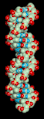 Spinning DNA generic model.
 -
 An oversimplified sketch of the double helix structure of A-DNA.
 -
 A model of DNA replication based on the double helix concept.
 -
 Animated, space-filling molecular model of the B-DNA double helix
 -
 A large-scale Crick-Watson DNA model shown in the Museum of Príncipe Felipe.
 -
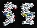
 Side view of molecular models of A-, B-, Z- DNA.
 -
 Oversimplified model of the A-DNA double helix.
 -
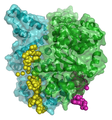
 Molecular modeling of RNA polymerase.
 -
 Molecular modeling of a bacterial DNA primase template.
 -
 Molecular modeling of DNA interactions with the carcinogen molecule MGMT.
 -
 3D Molecular model of DNA damaged by carcinogenic 2-aminofluorene(AF).
 -
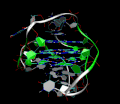
 Fig.6. Molecular modeling of DNA repair
 -
 Animated skeletal model of A-DNA.
 -
 Simplified models of chromatin.
 -
 Simplified model of chromosome structure.
 -
 A hypothetical quadruplex of guanine-rich DNA structures that may be involved in cancers.
 -
 3D Molecular Structure of the intramolecular human telomeric G-quadruplex in potassium solution.
 -
 DNA spacefilling molecular model
 -
 A model of a designed DNA tetrahedron.
 -
 15 m long DNA model, Naturalis Biodiversity Center


See also
- G-quadruplex
- Crystallography
- Crystal lattices
- Dinucleotide Property Database (DiProDB), designed to collect and analyse thermodynamic, structural and other dinucleotide traits
- X-ray microscopy
- X-ray scattering
- Neutron scattering
- Nucleic acid sequence
- Vibrational circular dichroism (VCD)
- Raman spectroscopy-microscopy and coherent anti-Stokes Raman spectroscopy (CARS)
- Sir Lawrence Bragg, FRS
- Comparison of nucleic acid simulation software
- AMBER
- CHARMM
- Abalone (molecular mechanics)
- Sirius visualization software
- QMC@Home
- NMR spectroscopy(FT-NMR)
- NMR imagingmicroscopy
- Microwave spectroscopy
- FT-infrared (IR)
- FT-near infrared spectroscopy(NIR)
- Spectral imaging, hyperspectral imaging, chemical imaging
- Fluorescence correlation spectroscopy
- Fluorescence cross-correlation spectroscopy and Förster resonance energy transfer (FRET)
- Confocal microscopy
- Obsolete models of DNA structure
References
- doi:10.1107/S0365110X53001940.)
{{cite journal}}: CS1 maint: multiple names: authors list (link - JSTOR 26060090.
- doi:10.1107/S0365110X52001635.)
{{cite journal}}: CS1 maint: multiple names: authors list (link - .
- S2CID 4253007.), .
{{cite journal}}: CS1 maint: multiple names: authors list (link - PMID 13168976.
- S2CID 4280080.)
{{cite journal}}: CS1 maint: multiple names: authors list (link - S2CID 36803326.
- PMID 14938364.
- PMID 14917668.
- ^ Chargaff E (1951). "Some recent studies on the composition and structure of nucleic acids". J Cell Physiol Suppl. 38 (Suppl).
- PMID 14778802.
- S2CID 2522535.
- S2CID 29369576.
- ^ Gautham, N. (25 May 2004). "Response to "Variety in DNA secondary structure"" (PDF). Current Science. 86 (10): 1352–1353. Retrieved 25 May 2012.
However, the discovery of topoisomerases took "the sting" out of the topological objection to the plectonaemic double helix. The more recent solution of the single crystal X-ray structure of the nucleosome core particle showed nearly 150 base pairs of the DNA (i.e. about 15 complete turns), with a structure that is in all essential respects the same as the Watson–Crick model. This dealt a death blow to the idea that other forms of DNA, particularly double helical DNA, exist as anything other than local or transient structures.
[dead link] - ^ Hosemann R., Bagchi R.N., Direct analysis of diffraction by matter, North-Holland Publs., Amsterdam – New York, 1962.
- .
- S2CID 43009840.
- .
- PMID 15600335.
- S2CID 29794525.
- PMID 16834438.
- PMID 17763481.
- PMID 17036134.
- ^ "Department of Physics, Cavendish Laboratory - Molecular Biophysics". Archived from the original on 23 May 2009. Retrieved 17 May 2009.
- ^ "Kryptowährungen und Physik – Planetphysics". Archived from the original on 31 March 2009. Retrieved 17 May 2009.
Further reading
- Applications of Novel Techniques to Health Foods, Medical and Agricultural Biotechnology.(June 2004) I. C. Baianu, P. R. Lozano, V. I. Prisecaru and H. C. Lin., q-bio/0406047.
- F. Bessel, Untersuchung des Theils der planetarischen Störungen, Berlin Abhandlungen (1824), article 14.
- Sir Lawrence Bragg, FRS. The Crystalline State, A General survey. London: G. Bells and Sons, Ltd., vols. 1 and 2., 1966., 2024 pages.
- Cantor, C. R. and Schimmel, P.R. Biophysical Chemistry, Parts I and II., San Francisco: W.H. Freeman and Co. 1980. 1,800 pages.
- Voet, D. and J.G. Voet. Biochemistry, 2nd Edn., New York, Toronto, Singapore: John Wiley & Sons, Inc., 1995, ISBN 0-471-58651-X., 1361 pages.
- Watson, G. N. A Treatise on the Theory of Bessel Functions., (1995) Cambridge University Press. ISBN 0-521-48391-3.
- Watson, James D. Molecular Biology of the Gene. New York and Amsterdam: W.A. Benjamin, Inc. 1965., 494 pages.
- Wentworth, W.E. Physical Chemistry. A short course., Malden ( Mass.): Blackwell Science, Inc. 2000.
- Herbert R. Wilson, FRS. Diffraction of X-rays by proteins, Nucleic Acids and Viruses., London: Edward Arnold (Publishers) Ltd. 1966.
- Kurt Wuthrich. NMR of Proteins and Nucleic Acids., New York, Brisbane, Chicester, Toronto, Singapore: J. Wiley & Sons. 1986., 292 pages.
- Hallin PF, David Ussery D (2004). "CBS Genome Atlas Database: A dynamic storage for bioinformatic results and DNA sequence data". Bioinformatics. 20 (18): 3682–6. PMID 15256401.
- Zhang CT, Zhang R, Ou HY (2003). "The Z curve database: a graphic representation of genome sequences". Bioinformatics. 19 (5): 593–599. PMID 12651717.
External links
- DNA the Double Helix Game From the official Nobel Prize website
- MDDNA: Structural Bioinformatics of DNA
- Double Helix 1953–2003 National Centre for Biotechnology Education
- DNAlive: a web interface to compute DNA physical properties. Also allows cross-linking of the results with the UCSC Genome browser and DNA dynamics.
- Further details of mathematical and molecular analysis of DNA structure based on X-ray data
- Bessel functions corresponding to Fourier transforms of atomic or molecular helices.[dead link]
- overview of STM/AFM/SNOM principles with educative videos
Databases for DNA molecular models and sequences
- X-ray diffraction
- NDB ID: UD0017 Database
- X-ray Atlas -database
- PDB files of coordinates for nucleic acid structures from X-ray diffraction by NA (incl. DNA) crystals
- Structure factors downloadable files in CIF format
- Neutron scattering
- X-ray microscopy
- Electron microscopy
- NMR databases
- NMR Atlas--database
- mmcif downloadable coordinate files of nucleic acids in solution from 2D-FT NMR data
- NMR constraints files for NAs in PDB format
- Genomic and structural databases
- CBS Genome Atlas Database — contains examples of base skews.
- The Z curve database of genomes — a 3-dimensional visualization and analysis tool of genomes.
- DNA and other nucleic acids' molecular models: Coordinate files of nucleic acids molecular structure models in PDB and CIF formats
- Atomic force microscopy
Types of nucleic acids | |||||||
|---|---|---|---|---|---|---|---|
| Constituents | |||||||
| Ribonucleic acids (coding, non-coding) |
| ||||||
| Deoxyribonucleic acids | |||||||
| Analogues | |||||||
| Cloning vectors | |||||||