
Movat's stain
Movat's stain is a pentachrome stain originally developed by Henry Zoltan Movat (1923–1995), a Hungarian-Canadian Pathologist in Toronto[1] in 1955 to highlight the various constituents of connective tissue, especially cardiovascular tissue, by five colors in a single stained slide.[2] In 1972, H. K. Russell, Jr. modified the technique so as to reduce the time for staining and to increase the consistency and reliability of the staining, creating the Russell–Movat stain.[3]
| Colour | Tissue type |
|---|---|
| Black | Nuclei; elastic fibres |
| Yellow | Collagen fibres; reticular fibres |
| Blue | Ground substance; mucin |
| Bright red | Fibrin |
| Red | Muscle |
Principle
Modified Russell–Movat
alcian blue
, Verhoeff
reticulin fibers are unstained by a reaction with phosphotungstic acid and stained in yellow by saffron
.
Uses
Modified Russell–Movat staining is used to study the
connective tissues. It can also be used to diagnose vascular and lung diseases.[5]
Gallery
-
 Movat's stain showing amyloid (brown) and fibrosis (yellow) of the heart
 -
 Movat's stain showing thickening of the spongiosa layer (blue) inmyxomatous degeneration of the aortic valve
 -
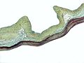
 Movat's stain showingcoronary artery atherosclerosis


References
- ^ Haust, M. Daria (April 1996). "In Memoriam: Dr. Henry Zoltan Movat, MD (Innsbruck), MSc, PhD (Queen's)" (PDF). Pathology News: Newsletter. Vol. 3, no. 4. Department of Pathology and Molecular Medicine, Queen's University. pp. 6–8. Retrieved 24 March 2023.
- PMID 13248341.
- PMID 4114784.
- Perelman School of Medicine, the University of Pennsylvania. Archived from the originalon 2012-08-04. Retrieved 2012-08-08.
- ^ "Modified Russel-Movat - Histalim". Archived from the original on 2016-08-10. Retrieved 2016-06-27.
See also
This article related to pathology is a stub. You can help Wikipedia by expanding it. |