
Mucormycosis
| Mucormycosis | |
|---|---|
| Other names | Zygomycosis, isavuconazole, posaconazole[8] |
| Prognosis | Poor[9] |
| Frequency | Rare[8] |
Mucormycosis, also known as black fungus, The fatality rate is about 54%.
It is spread by spores of
Diagnosis is by
Mucormycosis is usually rare,
Classification
Generally, mucormycosis is classified into five main types according to the part of the body affected.[14][23] A sixth type has been described as mucormycosis of the kidney,[1] or miscellaneous, i.e., mucormycosis at other sites, although less commonly affected.[23]
- kidney transplant.[14]
- organ transplant or a stem cell transplant.[14]
- Stomach and intestine (gastrointestinal); more common among young, premature, and low birth weight infants, who have had antibiotics, surgery, or medications that lower the body's ability to fight infection.[14]
- leukaemia, poorly controlled diabetes, graft-versus-host disease, HIV and intravenous drug use.[5][14]
- Widespread (disseminated); when the infection spreads to other organs via the blood.[14]
Signs and symptoms

Signs and symptoms of mucormycosis depend on the location in the body of the infection.[6] Infection usually begins in the mouth or nose and enters the central nervous system via the eyes.[5]
If the fungal infection begins in the nose or sinus and extends to brain, symptoms and signs may include one-sided eye pain or
Fever, cough, chest pain, and
Invasion of the
Cause
Mucormycosis is a
The fungal spores are present in the environment, can be found on items such as moldy bread and fruit, and are breathed in frequently, but cause disease only in some people.
Risk factors
Predisposing factors for mucormycosis include
Corticosteroids are commonly used in the treatment of COVID-19 and reduce damage caused by the body's own immune response to the virus. They are immunosuppressant and increase blood sugar levels in both diabetic and non-diabetic patients. It is thought that both these effects may contribute to cases of mucormycosis.[34][35][21]
Mechanism
Most people are frequently exposed to Mucorales without developing the disease.[31] Mucormycosis is generally spread by breathing in, eating food contaminated by, or getting spores of molds of the Mucorales type in an open wound.[15] It is not transmitted between people.[14]
The precise mechanism by which diabetics become susceptible is unclear. In vivo, a high sugar level alone does not permit the growth of the fungus, but acidosis alone does.[1][7] People with high sugar levels frequently have high iron levels, also known to be a risk factor for developing mucormycosis.[7] In people taking deferoxamine, the iron removed is captured by siderophores on Rhizopus species, which then use the iron to grow.[36]
Diagnosis
There is no blood test that can confirm the diagnosis.
Imaging
Culture and biopsy
To confirm the diagnosis, biopsy samples can be cultured.[13][37] Culture from biopsy samples does not always give a result as the organism is very fragile.[17] Microscopy can usually determine the genus and sometimes the species, but may require an expert mycologist.[17] The appearance of the fungus under the microscope can vary but generally shows wide (10–20 micron), ribbon-like filaments that generally do not have septa and that—unlike in aspergillosis—branch at right angles, resembling antlers of a moose, which may be seen to be invading blood vessels.[12][37]
-
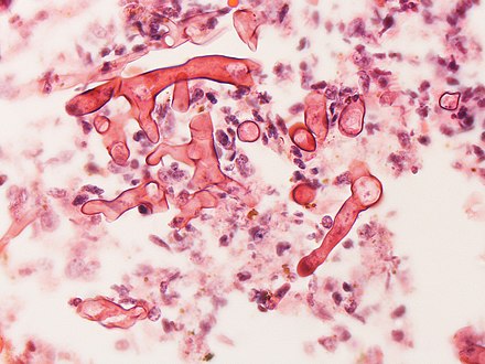 Ribbon-like hyphae which branch at 90°
 -
 Hyphae in blood vessel
 -
![Mature sporangium of a Mucor[40]](//upload.wikimedia.org/wikipedia/commons/thumb/c/c5/Mature_sporangium_of_a_Mucor_sp._fungus.jpg/483px-Mature_sporangium_of_a_Mucor_sp._fungus.jpg) Mature sporangium of a Mucor[40]
Mature sporangium of a Mucor[40]

.jpg)
![Mature sporangium of a Mucor[40]](/File:Mature_sporangium_of_a_Mucor_sp._fungus.jpg)
Other
Differential diagnosis
Other filamentous fungi may however look similar.
Prevention
Preventive measures include wearing a face mask in dusty areas, washing hands, avoiding direct contact with water-damaged buildings, and protecting skin, feet, and hands where there is exposure to soil or manure, such as gardening or certain outdoor work.
Treatment
Treatment involves a combination of
Medication
Once mucormycosis is suspected,
Surgery
Surgery can be very drastic, and, in some cases of disease involving the nasal cavity and the brain, removal of infected brain tissue may be required. Removal of the palate, nasal cavity, or eye structures can be very disfiguring.[26] Sometimes more than one operation is required.[31]
Other considerations
The disease must be monitored carefully for any signs of reemergence.
Prognosis
It tends to progress rapidly and is fatal in about half of sinus cases, two thirds of lung cases, and almost all cases of the widespread type.
As treatment usually requires extensive and often disfiguring facial surgery, the
Epidemiology
The true incidence and prevalence of mucormycosis may be higher than appears.[36] Mucormycosis is rare, affecting fewer than 1.7 people per million population each year in San Francisco.[8][46] It is around 80 times more prevalent in India, where it is estimated that there are around 0.14 cases per 1000 population,[19] and where its incidence has been rising.[47] Causative fungi are highly dependent on location. Apophysomyces variabilis has its highest prevalence in Asia and Lichtheimia spp. in Europe.[20] It is the third most common serious fungal infection to infect people, after aspergillosis and candidiasis.[48]
Diabetes is the main underlying disease in low and middle-income countries, whereas, blood cancers and organ transplantation are the more common underlying problems in developed countries.[19] As new immunomodulating drugs and diagnostic tests are developed, the statistics for mucormycosis have been changing.[19] In addition, the figures change as new genera and species are identified, and new risk factors reported such as tuberculosis and kidney problems.[19]
COVID-19–associated mucormycosis
_cases.png)
During the
Recurrence of mucormycosis during COVID-19 second wave in India
Pre-COVID mucormycosis was a very rare infection, even in India. It is so rare that an ENT (ear, nose, throat) doctor would not witness often a case during their university time. So, the documentation available on the treatment of mucormycosis is limited. In fact, there used to be a couple of mucormycosis expert ENT surgeons for millions of people pre-pandemic. The sudden rise in mucormycosis cases has left a majority of the ENT doctors with no option but to accept mucormycosis cases, as the expert doctors were very much occupied and the patient would die if left untreated. The majority of the ENT doctors had to manage with minimal or no experience on mucormycosis, this has led to the recurrence of mucormycosis in the patients they treated. When a highly experienced doctor in mucormycosis treats a patient even he cannot guarantee that the individual is completely cured and will not have a relapse of mucormycosis; an inexperienced ENT surgeon will definitely have a high number of patients with recurrence due to which there were many recurrent cases of mucormycosis although it did not get the limelight of media or the Indian Government.
History
The first case of mucormycosis was possibly one described by Friedrich Küchenmeister in 1855.[1] Fürbringer first described the disease in the lungs in 1876.[52] In 1884, Lichtheim established the development of the disease in rabbits and described two species; Mucor corymbifera and Mucor rhizopodiformis, later known as Lichtheimia and Rhizopus, respectively.[1] In 1943, its association with poorly controlled diabetes was reported in three cases with severe sinus, brain and eye involvement.[1]
In 1953,
Naming
Arnold Paltauf coined the term "Mycosis Mucorina" in 1885, after describing a case with systemic symptoms involving the sinus, brain and gastrointestinal tract, following which the term "mucormycosis" became popular.
COVID-19–associated mucormycosis
COVID-19 associated mucormycosis cases were reported during first and second(delta) wave, with maximum number of cases in delta wave.[11] There were no cases reported during the Omicron wave.[11] A number of cases of mucormycosis, aspergillosis, and candidiasis, linked to immunosuppressive treatment for COVID-19 were reported during the COVID-19 pandemic in India in 2020 and 2021.[4][39] One review in early 2021 relating to the association of mucormycosis and COVID-19 reported eight cases of mucormycosis; three from the U.S., two from India, and one case each from Brazil, Italy, and the UK.[21] The most common underlying medical condition was diabetes.[21] Most had been in hospital with severe breathing problems due to COVID-19, had recovered, and developed mucormycosis 10–14 days following treatment for COVID-19. Five had abnormal kidney function tests, three involved the sinus, eye and brain, three the lungs, one the gastrointestinal tract, and in one the disease was widespread.[21] In two of the seven deaths, the diagnosis of mucormycosis was made at postmortem.[21] That three had no traditional risk factors led the authors to question the use of steroids and immunosuppressive drugs.[21] Although, there were cases without diabetes or use of immunosuppressive drugs. There were cases reported even in children.[11] In May 2021, the BBC reported increased cases in India.[34] In a review of COVID-19-related eye problems, mucormycosis affecting the eyes was reported to occur up to several weeks following recovery from COVID-19.[39] It was observed that people with COVID-19 were recovering from mucormycosis a bit easily when compared to non-COVID-19 patients. This is because unlike non-COVID-19 patients with severe diabetes, cancer or HIV, the recovery time required for the main cause of immune suppression is temporary.[11]
Other countries affected included Pakistan,[54] Nepal,[55] Bangladesh,[56] Russia,[57] Uruguay,[58] Paraguay,[59] Chile,[60] Egypt,[61] Iran,[62] Brazil,[63] Iraq,[64] Mexico,[65] Honduras,[66] Argentina[67] Oman,[68] and Afghanistan.[69] One explanation for why the association has surfaced remarkably in India is high rates of COVID-19 infection and high rates of diabetes.[70] In May 2021, the Indian Council of Medical Research issued guidelines for recognising and treating COVID-19–associated mucormycosis.[71] In India, as of 28 June 2021, over 40,845 people have been confirmed to have mucormycosis, and 3,129 have died. From these cases, 85.5% (34,940) had a history of being infected with SARS-CoV-2 and 52.69% (21,523) were on steroids, also 64.11% (26,187) had diabetes.[72][73]
Society and culture
The disease has been reported in natural disasters and catastrophes;
In 2014, details of a lethal mucormycosis outbreak that occurred in 2008 emerged after television and newspaper reports responded to an article in a pediatric medical journal.[77][78] Contaminated hospital linen was found to be spreading the infection. A 2018 study found many freshly laundered hospital linens delivered to U.S. transplant hospitals were contaminated with Mucorales.[79] Another study attributed an outbreak of hospital-acquired mucormycosis to a laundry facility supplying linens contaminated with Mucorales. The outbreak stopped when major changes were made at the laundry facility. The authors raised concerns on the regulation of healthcare linens.[80]
Other animals
Mucormycosis in other animals is similar, in terms of frequency and types, to that in people.[81] Cases have been described in cats, dogs, cows, horses, dolphins, bison, and seals.[81]
References
- ^ ISBN 978-93-86261-83-0.
- ^ a b "Orphanet: Zygomycosis". www.orpha.net. Archived from the original on May 13, 2021. Retrieved May 13, 2021.
- ^ PMID 33985993.
- ^ ISBN 978-93-90486-29-8.
- ^ ISBN 978-1-4419-1577-1.
- ^ a b c d e f g "Symptoms of Mucormycosis". www.cdc.gov. January 14, 2021. Retrieved May 25, 2021.
- ^ PMID 16020690.
- ^ a b c d e f g h i "Mucormycosis". NORD (National Organization for Rare Disorders). Archived from the original on May 26, 2021. Retrieved May 25, 2021.
- ^ PMID 31335084.
- ^ a b c d e "People at Risk For Mucormycosis and prevention". www.cdc.gov. February 2, 2021. Retrieved May 25, 2021.
- ^ a b c d e f Meghanadh KR (May 15, 2021). "Mucormycosis / Black fungus infection". Medy Blog. Retrieved June 27, 2021.
- ^ ISBN 978-0-7020-6830-0.
- ^ a b "ICD-11 - ICD-11 for Mortality and Morbidity Statistics". icd.who.int. Retrieved May 25, 2021.
- ^ a b c d e f g h i j "About Mucormycosis". www.cdc.gov. May 25, 2021.
- ^ S2CID 210984392.
- ^ "Where Mucormycosis Comes From". www.cdc.gov. February 1, 2021. Retrieved May 25, 2021.
- ^ ISBN 978-0-12-820703-1.
- ^ a b "Mucormycosis Statistics | Mucormycosis | Fungal Diseases | CDC". www.cdc.gov. May 5, 2020. Retrieved May 25, 2021.
- ^ PMID 33147877.
- ^ PMID 31877973.
- ^ PMID 33544266.
- PMID 34192610.
- ^ S2CID 22454217.
- ^ a b McDonald PJ. "Mucormycosis (Zygomycosis) Clinical Presentation: History and Physical Examination". emedicine.medscape.com. Retrieved May 28, 2021.
- ISBN 0-632-04568-X.
- ^ a b c "MedlinePlus Medical Encyclopedia: Mucormycosis". Retrieved May 19, 2008.
- PMID 24020743.
- ^ PMID 16020690.
- ISBN 978-1-55581-958-3.
- PMID 32811466.
- ^ a b c d e f g h McDonald PJ (September 10, 2018). "Mucormycosis (Zygomycosis): Background, Etiology and Pathophysiology, Epidemiology". Medscape.
- ^ a b c "For Healthcare Professionals | Mucormycosis | CDC". www.cdc.gov. June 17, 2020. Retrieved May 25, 2021.
- PMID 35357212.
- ^ a b Biswas S (May 9, 2021). "Mucormycosis: The 'black fungus' maiming Covid patients in India". BBC News. British Broadcasting Corporation. Retrieved May 11, 2021.
- PMID 33333012.
- ^ PMID 30901907.
- ^ a b c d e f g h McDonald PJ. "Mucormycosis (Zygomycosis) Workup: Approach Considerations, Laboratory Tests, Radiologic Studies". emedicine.medscape.com. Retrieved May 25, 2021.
- ^ "Diagnosis and Testing of Mucormycosis | Mucormycosis | CDC". www.cdc.gov. January 14, 2021.
- ^ PMID 33595463.
- ^ "Details - Public Health Image Library(PHIL)". phil.cdc.gov. Retrieved May 26, 2021.
- ^ PMID 31699664. (several authors)
- ^ McDonald PJ. "Mucormycosis (Zygomycosis) Differential Diagnoses". emedicine.medscape.com. Retrieved May 25, 2021.
- ISBN 978-0-85711-369-6.
- ^ McDonald PJ (September 10, 2018). "What is the role of isavuconazole (Cresemba) in the treatment of mucormycosis (zygomycosis)?". www.medscape.com. Retrieved May 25, 2021.
- ^ Rebecca J. Frey. "Mucormycosis". Health A to Z. Archived from the original on May 18, 2008. Retrieved May 19, 2008.
- ^ "Mucormycosis Statistics | Mucormycosis | Fungal Diseases | CDC". www.cdc.gov. June 5, 2020. Archived from the original on May 21, 2021. Retrieved May 22, 2021.
- ISBN 978-0-323-41649-8.
- PMID 22247442.
- ^ Schwartz I, Chakrabarti A (June 2, 2021). "'Black fungus' is creating a whole other health emergency for Covid-stricken India". The Guardian. Retrieved June 3, 2021.
- ^ Wadhawan DW, Jain P, Shrivastava S, Kunal K (May 19, 2021). "Rajasthan declares black fungus an epidemic; cases pile up in several states | 10 points". India Today. Retrieved May 20, 2021.
- ^ "Black fungus in India: Concern over drug shortage as cases rise". BBC News. May 19, 2021. Retrieved July 8, 2021.
- ^ PMID 29404149.
- PMID 13405736.
- ^ "'Cases of Black Fungus emerge across Pakistan'". The News International. May 12, 2021.
- ^ "Focused COVID-19 Media Monitoring, Nepal (May 24, 2021)". ReliefWeb. May 24, 2021.
- ^ "Bangladesh reports 1st death by black fungus". Anadolu Agency. May 25, 2021.
- ^ "Russia Confirms Rare, Deadly 'Black Fungus' Infections Seen in India". The Moscow Times. May 17, 2021.
- ^ "Paciente con COVID-19 se infectó con el "hongo negro"". EL PAÍS Uruguay (in Spanish). May 25, 2021.
- ^ "Confirman dos casos de "hongo negro" en Paraguay". RDN (in Spanish). May 27, 2021.
- ^ "Detectan primer caso de "hongo negro" en Chile en paciente con Covid-19: es el segundo reportado en Latinoamérica". El Mostrador (in Spanish). May 28, 2021.
- ^ "Mansoura University Hospital reports black fungus cases". Egypt Independent. May 28, 2021.
- ^ "Coronavirus in Iran: Power outages, black fungus, and warnings of a fifth surge". Track Persia. May 29, 2021. Archived from the original on May 29, 2021. Retrieved June 1, 2021.
- ^ "Casos suspeitos de fungo preto são investigados no Brasil; entenda". Catraca Livre (in Portuguese). May 31, 2021.
- ^ "Iraq detects five cases of the deadly "black fungus" among coronavirus patients". Globe Live Media. June 1, 2021.
- ^ "Detectan en Edomex posible primer caso de hongo negro en México". Uno TV (in Spanish). June 3, 2021.
- ^ "Salud confirma primer caso de hongo negro en Honduras". Diario El Heraldo (in Spanish). June 7, 2021.
- ^ ""Hongo negro": advierten que hay que estar atentos a la coinfección fúngica en pacientes con covid". Clarín (in Spanish). June 16, 2021.
- ^ "'Black fungus' detected in 3 COVID-19 patients in Oman". Al Jazeera. June 15, 2021.
- ^ "Afghanistan finds deadly 'black fungus' in virus patients – latest updates". TRT World. Retrieved July 17, 2021.
- ^ Runwal P (May 14, 2021). "A rare black fungus is infecting many of India's COVID-19 patients—why?". National Geographic. Archived from the original on May 14, 2021.
- ^ "ICMR releases diagnosis and management guidelines for COVID-19-associated Mucormycosis". Firstpost. May 17, 2021.
- ^ "India reports 40,854 cases of black fungus so far". Mint. June 28, 2021. Retrieved July 16, 2021.
- ^ "Delhi has more black fungus infections than active Covid-19 cases: Govt data". Mint. July 14, 2021. Retrieved July 16, 2021.
- ^ Fanfair, Robyn Neblett; et al. (July 29, 2011). "Notes from the Field: Fatal Fungal Soft-Tissue Infections After a Tornado – Joplin, Missouri, 2011". MMWR Weekly. 60 (29): 992.
- ^ Williams, Timothy (June 10, 2011) Rare Infection Strikes Victims of a Tornado in Missouri. New York Times.
- PMID 23215557.
- ^ Catalanello, Rebecca (April 16, 2014). "Mother believes her newborn was the first to die from fungus at Children's Hospital in 2008". NOLA.com.
- ^ "5 Children's Hospital patients died in 2008, 2009 after contact with deadly fungus".
We acknowledge that Children's Hospital is Hospital A in an upcoming article in The Pediatric Infectious Disease Journal. The safety and well-being of our patients are our top priorities, so as soon as a problem was suspected, the State Health Department and CDC were notified and invited to assist in the investigation. The hospital was extremely aggressive in trying to isolate and then eliminate the source of the fungus.
- PMID 30299481.
- PMID 34282829.
- ^ PMID 29538732.
Further reading
- Cornely OA, Alastruey-Izquierdo A, Arenz D, Chen SC, Dannaoui E, Hochhegger B, et al. (December 2019). "Global guideline for the diagnosis and management of mucormycosis: an initiative of the European Confederation of Medical Mycology in cooperation with the Mycoses Study Group Education and Research Consortium". The Lancet. Infectious Diseases. 19 (12): e405–e421. PMID 31699664.