
Muscularis mucosae
| Muscularis mucosae | |
|---|---|
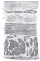
 Section of duodenum of cat. X 60. (Muscularis mucosae labeled at right, third from the top.) | |
| Details | |
| Identifiers | |
| Latin | lamina muscularis mucosae |
| TA98 | A05.4.01.016 |
| FMA | 68413 |
| Anatomical terminology | |
The muscularis mucosae (or lamina muscularis mucosae) is a thin layer (
bladder
; as it is discontinuous, it should not be regarded as a true muscularis mucosae.
In the gastrointestinal tract, the term mucosa or mucous membrane refers to the combination of epithelium, lamina propria, and (where it occurs) muscularis mucosae.[1] The etymology suggests this, since the Latin names translate to "the mucosa's own special layer" (lamina propria mucosae) and "muscular layer of the mucosa" (lamina muscularis mucosae).
The muscularis mucosae is composed of several thin layers of
smooth muscle fibers oriented in different ways which keep the mucosal surface and underlying glands in a constant state of gentle agitation to expel contents of glandular crypts and enhance contact between epithelium and the contents of the lumen
.
Additional images
-
 Section of the human esophagus. Moderately magnified.
 -
 Section of mucous membrane of human stomach, near the cardiac orifice.
 -
 Section of mucous membrane of human rectum. X 60.
 -
 Layers of stomach wall
 -
 General structure of the gut wall showing the Muscularis mucosa.


References
- ^ H.G. Burkitt et al., Wheater's Functional Histology, 3rd ed.
Stacey E. Mills — Histology for Pathologists: 3rd (third) Edition, page 670.
ISBN 9780781762410
External links
- aplab[dead link] - BioWeb at University of Wisconsin System
- Histology image: 14_08 at the University of Oklahoma Health Sciences Center — "Lung"
- Nosek, Thomas M. "Section 6/6ch1/s6ch1_11". Essentials of Human Physiology. Archived from the original on 2016-03-24.
- Anatomy photo: digestive/mammal/system1/system4 - Comparative Organology at University of California, Davis - "Mammal, whole system (LM, Low)"
- UIUC Histology Subject 830