
Necrotizing enterocolitis
| Necrotizing enterocolitis | |
|---|---|
antibiotics, surgery[2] | |
| Prognosis | Risk of death 25%[1] |
Necrotizing enterocolitis (NEC) is a devastating intestinal disease that affects premature or very low birth weight infants.[4][1] Symptoms may include poor feeding, bloating, decreased activity, blood in the stool, vomiting of bile, multi-organ failure, and even death.[1][2]
The exact cause is unclear.
Prevention includes the use of
About 7% of those who are born prematurely develop NEC; however the odds of an infant developing this illness is directly related to the intensive care unit they are placed in.[11][12][13][4][2] Onset is typically in the first four weeks of life.[2] Among those affected, about 25% die.[1] The sexes are affected with equal frequency.[14] The condition was first described between 1888 and 1891.[14]
Signs and symptoms
The condition is typically seen in premature infants, and the timing of its onset is generally inversely proportional to the gestational age of the baby at birth (i.e., the earlier a baby is born, the later signs of NEC are typically seen).[15]
Initial symptoms include feeding intolerance and
Cause
The exact cause is unclear.[1] Several risk factors have been implicated:[17]
Maternal factors
- Acid-suppressing medications
- Chorioamnionitis
- Cocaine abuse
- In utero growth restriction
- Increased body mass index
- Intrahepatic cholestasis during pregnancy
- Lack of prenatal steroids
- Mode of delivery
- Placental abruption
- Pre-eclampsia
- Smoking
Main risk factors
- Low birth weight
- Prematurity
- Formula feeding (bovine based)
- Intestinal dysbiosis
Other risk factors
- Acute hypoxia
- Antibiotic exposure
- Blood transfusions
- Cardiac anomalies
- Neonatal anemia
- Poor intestinal perfusion
- Prolonged use of indomethacinfor patent ductus arteriosus closure
Diagnosis
Diagnosis is usually suspected clinically, but often requires the aid of diagnostic imaging, most commonly radiography, which can show the intestines and may show areas with death tissue or a bowel perforation.[18] Specific radiographic signs of NEC are associated with specific Bell's stages of the disease:[19]
- Bell's stage 1 (suspected disease):
- Mild systemic disease (apnea, lethargy,[20] slowed heart rate, temperature instability)
- Mild intestinal signs (abdominal distention, increased gastric residuals, bloody stools)
- Nonspecific or normal radiological signs
- Bell's stage 2 (definite disease):
- Mild to moderate systemic signs
- Additional intestinal signs (absent bowel sounds, abdominal tenderness)
- Specific radiologic signs (pneumatosis intestinalis or portal venous gas)
- Laboratory changes (metabolic acidosis, too few platelets in the bloodstream)
- Bell's stage 3 (advanced disease):
- Severe systemic illness (low blood pressure)
- Additional intestinal signs (striking abdominal distention, peritonitis)
- Severe radiologic signs (pneumoperitoneum)
- Additional laboratory changes (metabolic and respiratory acidosis, disseminated intravascular coagulation)
-
 Alimentary tract of infant showing intestinal necrosis, pneumatosis intestinalis, and perforation site (arrow) (autopsy)
 -
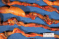 Close-up of intestine of infant showing necrosis and pneumatosis intestinalis (autopsy)


Diagnosis of NEC is more challenging in premature infants, due to inexplicit symptoms and radiographic signs. The most preterm infant is at highest risk of developing NEC.[23]
Prevention
Prevention includes the use of breast milk and probiotics.[2] A 2012 policy by the American Academy of Pediatrics recommended feeding preterm infants human milk, finding "significant short- and long-term beneficial effects," including reducing the rate of NEC by a factor of one-half to three-quarters.[24]
Small amounts of oral feeds of human milk starting as soon as possible, while the infant is being primarily fed intravenously, primes the immature gut to mature and become ready to receive greater intake by mouth.
Towards understanding intervention with human milk, experts have noted cow's and human milk differ in their immunoglobular and glycan compositions.[26][27] Due to their relative ease of production, human milk oligosaccharides (HMO) are a subject of particular interest in supplementation and intervention.[28]
A Cochrane review in 2020 (updated in 2023) found low- to moderate-quality evidence that supplementation of probiotics enterally "prevents severe NEC, as well as all-cause mortality in preterm infants" but cautioned that the evidence was not sufficient to inform policy and practice and that further high-quality trials are needed.[29]
Advancing
Treatment
If a baby is diagnosed with NEC, treatment should begin immediately.[18] Treatment consists primarily of supportive care, including providing bowel rest by stopping enteral feeds, gastric decompression with intermittent suction, fluid repletion to correct electrolyte abnormalities and third-space losses, support for blood pressure, parenteral nutrition,[32] and prompt antibiotic therapy.
Monitoring is clinical, although serial supine and left lateral decubitus abdominal X-rays should be performed every six hours.[33]
As an infant recovers from NEC, feeds are gradually introduced. "Trophic feeds" or low-volume feeds (<20 ml/kg/day) are usually initiated first. How and what to feed are determined by the extent of
Where the disease is not halted through medical treatment alone, or when the
In the case of an infant whose bowel is left in discontinuity, the surgical creation of a mucous
Prognosis
Typical recovery from NEC if medical, nonsurgical treatment succeeds, includes 10–14 days or more without oral intake, and then demonstrated ability to resume feedings and gain weight. Recovery from NEC alone may be compromised by co-morbid conditions that frequently accompany prematurity. Long-term complications of medical NEC include bowel obstruction and anemia.[citation needed]
In the United States, NEC caused 355 deaths per 100,000 live births in 2013, down from 484 per 100,000 live births in 2009. Rates of death were almost three times higher for the black population than for the white population.[35]
When NEC is diagnosed and treated immediately, the prognosis for babies is generally very good. Most babies recover fully without any additional health problems.[18] Overall, about 70-80% of infants who develop NEC survive.[36] Medical management of NEC shows an increased chance of survival compared to surgical management.[36] Despite a significant mortality risk, long-term prognosis for infants undergoing NEC surgery is improving, with survival rates of 70–80%. However, "Surgical NEC" survivors are still at risk for possible long-term complications, such as narrowing of the intestines[18] or short bowel syndrome and neurodevelopmental disability.
Society and advocacy
The NEC Society is a 501(c)(3), non-profit organization dedicated to building a world without necrotizing enterocolitis (NEC) through research, advocacy, and education. The NEC Society was launched in January 2014 by Jennifer Canvasser after her son, Micah, died from complications of NEC just before his first birthday. The NEC Society is a patient-led organization that collaborates with expert clinicians and researchers to better understand, prevent, and treat this devastating neonatal intestinal disease. Today, patient-families and experts from around the world work together to improve outcomes for the most vulnerable infants at risk of NEC. Their work and numerous initiatives combine the patient-family perspective with solutions based on the best available scientific evidence.[citation needed]
NEC Symposium
The NEC Society hosts an in-person, biennial Symposium where clinicians, scientists and patient-families come together to listen, learn and collaborate. It is held as an “All-In Meeting", where all stakeholders are fully integrated and empowered. Patient-families are central to the planning, preparation, and execution of the meeting. Each session is dedicated to a baby affected by NEC. Patient-families take part in each session as faculty and also present posters.[citation needed]
References
- ^ a b c d e f g h i j k "Necrotizing Enterocolitis – Pediatrics – Merck Manuals Professional Edition". Merck Manuals Professional Edition. February 2017. Retrieved 12 December 2017.
- ^ S2CID 39251333.
- ISBN 9780781737708.
- ^ a b Gephart S.M., Quinn M. A call to action to fight for equity and end necrotizing enterocolitis disparities. Adv. Neonatal Care. 2021;21(5):333-335. doi:10.1097/ANC.0000000000000940
- PMID 30115546.
- PMID 28351838.
- PMID 22920508.
- PMID 27543379.
- PMID 2056401.
- S2CID 17281723.
- ^ Gephart SM, Spitzer AR, Effken JA, Dodd E, Halpern M, McGrath JM. Discrimination of GutCheckNEC: a clinical risk index for necrotizing enterocolitis. J Perinatol. 2014;34(6):468-475.
- ^ Horbar JD, Edwards EM, Greenberg LT, et al. Variation in performance of neona-tal intensive care units in the United States. JAMA Pediatr. 2017;171(3):e164396.
- ^ Uauy RD, Fanaroff AA, Korones SB, Phillips EA, Phillips JB, Wright LL. Necrotizing enterocolitis in very low birth weight infants: biodemographic and clinical corre-lates. National Institute of Child Health and Human Development Neonatal Research Network. J Pediatr. 1991;119(4):630-638.
- ^ S2CID 29437889.
- S2CID 26079047.
- ^ "Necrotizing Enterocolitis". The Lecturio Medical Concept Library. Retrieved 11 August 2021.
- PMID 30740215.
- ^ a b c d Cline M (2021-07-14). "Necrotizing Enterocolitis: A Guide for Preemie Parents". Birth Injury Guide. Retrieved 2021-08-19.
- S2CID 37235496.
- ^ Schanler RJ, Abrams SA, Kim MS (2016). "Clinical features and diagnosis of necrotizing enterocolitis in newborns". UpToDate.
- ^ Muchantef K, Epelman M, Darge K, Kirpalani H, Laje P, Anupindi SA. Sonographic and radiographic imaging features of the neonate with necrotizing enterocolitis: correlating findings with outcomes. Pediatr Radiol. 2013 Jun 15.
- ISBN 978-0-7817-8251-7.
- S2CID 38176277.
- PMID 22371471.
Meta-analyses of four randomized clinical trials performed over the period 1983 to 2005 support the conclusion that feeding preterm infants human milk is associated with a significant reduction (58%) in the incidence of NEC. A more recent study of preterm infants fed an exclusive human milk diet compared with those fed human milk supplemented with cow's milk-based infant formula products noted a 77% reduction in NEC.
- S2CID 36737314.
- PMID 22513036.
- PMID 31111150.
- PMID 27634978.
- PMID 37493095.
- PMID 34427330.
- PMID 35049036.
- PMID 8070233.
- PMID 21247316.
- ^ S2CID 49419886.
- PMID 26905861.
- ^ a b Schanler RJ, Abrams SA, Kim MS. Management of necrotizing enterocolitis in newborns. UpToDate (Report).