
Nephron
| Nephron | |
|---|---|
Metanephric blastema (intermediate mesoderm) | |
| System | Urinary system |
| Identifiers | |
| MeSH | D009399 |
| FMA | 17640 |
| Anatomical terminology] | |
The nephron is the minute or microscopic structural and functional unit of the
The interior of Bowman's capsule, called Bowman's space, collects the filtrate from the filtering capillaries of the
The four mechanisms used to create and process the filtrate (the result of which is to convert blood to urine) are
Some diseases of the nephron predominantly affect either the glomeruli or the tubules. Glomerular diseases include diabetic nephropathy, glomerulonephritis and IgA nephropathy; renal tubular diseases include acute tubular necrosis and polycystic kidney disease.
Structure

The nephron is the functional unit of the kidney.[2] This means that each separate nephron is where the main work of the kidney is performed.
A nephron is made of two parts:
- a renal corpuscle, which is the initial filtering component, and
- a renal tubule that processes and carries away the filtered fluid.[3]: 1024
Renal corpuscle

B. Glomerular basement membrane: 1. lamina rara interna 2. lamina densa 3. lamina rara externa
C. Podocytes: 1. enzymatic and structural proteins 2. filtration slit 3. diaphragm
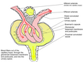
The renal corpuscle is the site of the filtration of blood plasma. The renal corpuscle consists of the glomerulus, and the glomerular capsule or Bowman's capsule.[3]: 1027
The renal corpuscle has two poles: a vascular pole and a tubular pole.[4]: 397 The arterioles from the renal circulation enter and leave the glomerulus at the vascular pole. The glomerular filtrate leaves the Bowman's capsule at the renal tubule at the urinary pole.
Glomerulus
The glomerulus is the network known as a tuft, of filtering capillaries located at the vascular pole of the renal corpuscle in Bowman's capsule. Each glomerulus receives its blood supply from an afferent arteriole of the renal circulation. The glomerular blood pressure provides the driving force for water and solutes to be filtered out of the blood plasma, and into the interior of Bowman's capsule, called Bowman's space.
Only about a fifth of the plasma is filtered in the glomerulus. The rest passes into an efferent arteriole. The diameter of the efferent arteriole is smaller than that of the afferent, and this difference increases the hydrostatic pressure in the glomerulus.
Bowman's capsule
The Bowman's capsule, also called the glomerular capsule, surrounds the glomerulus. It is composed of a visceral inner layer formed by specialized cells called podocytes, and a parietal outer layer composed of simple squamous epithelium. Fluids from blood in the glomerulus are ultrafiltered through several layers, resulting in what is known as the filtrate.
The filtrate next moves to the renal tubule, where it is further processed to form urine. The different stages of this fluid are collectively known as the tubular fluid.
Renal tubule
The renal tubule is long pipe like structure containing the tubular fluid filtered through the glomerulus.[5] After passing through the renal tubule, the filtrate continues to the collecting duct system.[6]
The components of the renal tubule are:
- Proximal convoluted tubule (lies in cortex and lined by simple cuboidal epithelium with brush borderswhich help to increase the area of absorption greatly.)
- Loop of Henle (hair-pin like, i.e. U-shaped, and lies in medulla)
- Descending limb of loop of Henle
- Ascending limb of loop of Henle
- The ascending limb of loop of Henle is divided into 2 segments: Lower end of ascending limb is very thin and is lined by simple squamous epithelium. The distal portion of ascending limb is thick and is lined by simple cuboidal epithelium.
- Thin ascending limb of loop of Henle
- Thick ascending limb of loop of Henle(enters cortex and becomes the distal convoluted tubule.)
- Distal convoluted tubule
- Collecting tubule
The epithelial cells that form these nephron segments can be distinguished by the shapes of their actin cytoskeleton.[7]
Blood from the efferent arteriole, containing everything that was not filtered out in the glomerulus, moves into the peritubular capillaries, tiny blood vessels that surround the loop of Henle and the proximal and distal tubules, where the tubular fluid flows. Substances then reabsorb from the latter back to the blood stream.
The peritubular capillaries then recombine to form an efferent venule, which combines with efferent venules from other nephrons into the renal vein, and rejoins the main bloodstream.
Length difference
Cortical nephrons (the majority of nephrons) start high in the cortex and have a short loop of Henle which does not penetrate deeply into the medulla. Cortical nephrons can be subdivided into superficial cortical nephrons and midcortical nephrons.[8]
Juxtamedullary nephrons[
Juxtamedullary nephrons are found only in birds and mammals, and have a specific location: medullary refers to the
The juxtamedullary nephrons comprise only about 15% of the nephrons in the human kidney.[1]: 24 However, it is this type of nephron which is most often depicted in illustrations of nephrons.
In humans, cortical nephrons have their renal corpuscles in the outer two thirds of the cortex, whereas juxtamedullary nephrons have their corpuscles in the inner third of the cortex.[1]: 24
Functions

The nephron uses four mechanisms to convert blood into urine: filtration, reabsorption, secretion, and excretion.[4]: 395–396 These apply to numerous substances. The structure and function of the epithelial cells lining the lumen change during the course of the nephron, and have segments named by their location and which reflects their different functions.

Proximal tubule
The proximal tubule as a part of the nephron can be divided into an initial convoluted portion and a following straight (descending) portion.[11] Fluid in the filtrate entering the proximal convoluted tubule is reabsorbed into the peritubular capillaries, including 80% of glucose, more than half of the filtered salt, water and all filtered organic solutes (primarily glucose and amino acids).[4]: 400–401
Loop of Henle
The loop of Henle is a U-shaped tube that extends from the proximal tubule. It consists of a descending limb and an ascending limb. It begins in the cortex, receiving filtrate from the proximal convoluted tubule, extends into the medulla as the descending limb, and then returns to the cortex as the ascending limb to empty into the distal convoluted tubule. The primary role of the loop of Henle is to enable an organism to produce concentrated urine, not by increasing the tubular concentration, but by rendering the interstitial fluid hypertonic.[1]: 67
Considerable differences aid in distinguishing the descending and ascending limbs of the loop of Henle. The
Unlike the descending limb, the
Distal convoluted tubule
The
Connecting tubule
A part of Distal nephron. This is the final segment of the tubule before it enters the collecting duct system. Water, some salts and nitrogenous waste like urea and creatinine are passed out to collecting tubule.
Collecting duct system

Each distal convoluted tubule delivers its filtrate to a
Because it has a different origin during the
Though the collecting duct is normally impermeable to water, it becomes permeable in the presence of

Lower portions of the collecting organ are also permeable to urea, allowing some of it to enter the medulla, thus maintaining its high concentration (which is very important for the nephron).[1]: 73–74
Urine leaves the medullary collecting ducts through the
: 406–407Juxtaglomerular apparatus
The
Clinical significance
Patients in early stages of chronic kidney disease show an approximate 50% reduction in the number of nephrons, comparable to the nephron loss that occurs with aging (between ages 18–29 and 70–75).[13]
Diseases of the nephron predominantly affect either the glomeruli or the tubules. Glomerular diseases include diabetic nephropathy, glomerulonephritis and IgA nephropathy; renal tubular diseases include acute tubular necrosis, renal tubular acidosis, and polycystic kidney disease.
Additional images
-

-
 Glomerulus is red; Bowman's capsule is white.
 -
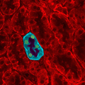 Kidney tissue
 -
 Glomerulus
 -
 This image shows the types of cells present in the glomerulus part of a kidney nephron. Podocytes, Endothelial cells, and Glomerular mesangial cell are present.


See also
References
- ^ a b c d e f g h Lote CJ (2012). Principles of Renal Physiology (5th ed.). Springer.
- ISBN 978-0-19-856878-0.
- ^ OCLC 192027371.
- ^ ISBN 978-0-07-184268-6.
- ^ "The Kidney Tubule I: Urine Production". Ecology & Evolutionary Biology - University of Colorado at Boulder. Archived from the original on October 2, 2007. Retrieved March 6, 2007.
- ISBN 0-88167-885-6.
- PMID 31605441.
- ^ Nosek TM. "Section 7/7ch03/7ch03p16". Essentials of Human Physiology. Archived from the original on 2016-03-24.
- ISBN 978-0-07-166339-7.
- ^ "Regulation of Urine Concentration". Anatomy & Physiology. CliffsNotes. Archived from the original on 25 October 2012. Retrieved 27 November 2012.
- ISBN 978-1-4160-2328-9.
- ^ Mitchell B, Sharma R (2009). Embriology (2nd ed.). Churchill Livingstone Elsevier.
- S2CID 53872296.
| Authority control databases: National |
|---|