Neuromelanin

Neuromelanin (NM) is a dark
Physical properties and structure

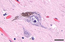
Neuromelanin gives specific brain sections, such as the substantia nigra or the locus coeruleus, distinct color. It is a type of melanin and similar to other forms of peripheral melanin. It is insoluble in organic compounds, and can be labeled by
Synthetic pathways
Neuromelanin is directly biosynthesized from
Neuromelanin biosynthesis is driven by excess
Function
Neuromelanin is found in higher concentrations in humans than in other
].Role in disease
Neuromelanin-containing
Neuromelanin has been shown to bind neurotoxic and toxic metals that could promote neurodegeneration.[5]
History
Dark pigments in the substantia nigra were first described in 1838 by Purkyně,[8] and the term neuromelanin was proposed in 1957 by Lillie,[9] though it has been thought to serve no function until recently. It is now believed to play a vital role in preventing cell death in certain parts of the brain. It has been linked to Parkinson's disease and because of this possible connection, neuromelanin has been heavily researched in the last decade.[10]
