
Node of Ranvier
| Node of Ranvier | |
|---|---|
 Drawing of a peripheral nerve axon (labeled "axis cylinder"), showing a node of Ranvier along with other features | |
 Nodes of Ranvier | |
| Details | |
| System | Nervous system |
| Location | Myelinated axon of a nerve |
| Identifiers | |
| Latin | incisura myelini |
| MeSH | D011901 |
| TH | H2.00.06.2.03015 |
| Anatomical terms of microanatomy | |
In
Overview

Many vertebrate axons are surrounded by a myelin sheath, allowing rapid and efficient
The internodal glial membranes are fused to form compact myelin, whereas the cytoplasm-filled paranodal loops of myelinating cells are spirally wrapped around the axon at both sides of the nodes. This organization demands a tight developmental control and the formation of a variety of specialized zones of contact between different areas of the myelinating cell membrane. Each node of Ranvier is flanked by paranodal regions where helicoidally wrapped glial loops are attached to the axonal membrane by a septate-like junction.
The segment between nodes of Ranvier is termed as the
Structure
The internodes are the myelin segments and the gaps between are referred to as nodes. The size and the spacing of the internodes vary with the fiber diameter in a curvilinear relationship that is optimized for maximal conduction velocity.[3] The size of the nodes span from 1–2 μm whereas the internodes can be up to (and occasionally even greater than)1.5 millimetres long, depending on the axon diameter and fiber type.
The structure of the node and the flanking paranodal regions are distinct from the internodes under the compact myelin sheath, but are very similar in CNS and PNS. The axon is exposed to the extra-cellular environment at the node and is constricted in its diameter. The decreased axon size reflects a higher packing density of
When a longitudinal section is made through a myelinating Schwann cell at the node, three distinctive segments are represented: the stereotypic internode, the paranodal region, and the node itself. In the internodal region, the Schwann cell has an outer collar of cytoplasm, a compact myelin sheath, and inner collar of cytoplasm, and the axolemma. At the paranodal regions, the paranodal cytoplasm loops contact thickenings of the axolemma to form septate –like junctions. In the node alone, the axolemma is contacted by several Schwann microvilli and contains a dense cytoskeletal undercoating.
Differences in the central and peripheral nervous systems
Although freeze fracture studies have revealed that the nodal axolemma in both the CNS and PNS is enriched in intra-membranous particles (IMPs) compared to the internode, there are some structural differences reflecting their cellular constituents.[4] In the PNS, specialized microvilli project from the outer collar of Schwann cells and come very close to nodal axolemma of large fibers. The projections of the Schwann cells are perpendicular to the node and are radiating from the central axons. However, in the CNS, one or more of the astrocytic processes come in close vicinity of the nodes. Researchers declare that these processes stem from multi-functional astrocytes, as opposed to from a population of astrocytes dedicated to contacting the node. On the other hand, in the PNS, the basal lamina that surrounds the Schwann cells is continuous across the node.
Composition
The nodes of Ranvier Na+/Ca2+ exchangers and high density of voltage-gated Na+ channels that generate action potentials. A sodium channel consists of a pore-forming α subunit and two accessory β subunits, which anchor the channel to extra-cellular and intra-cellular components. The nodes of Ranvier in the central and peripheral nervous systems mostly consist of αNaV1.6 and β1 subunits.
Molecular organization
The molecular organization of the nodes corresponds to their specialized function in impulse propagation. The level of sodium channels in the node versus the internode suggests that the number IMPs corresponds to sodium channels. Potassium channels are essentially absent in the nodal axolemma, whereas they are highly concentrated in the paranodal axolemma and Schwann cell membranes at the node.[4] The exact function of potassium channels have not quite been revealed, but it is known that they may contribute to the rapid repolarization of the action potentials or play a vital role in buffering the potassium ions at the nodes. This highly asymmetric distribution of voltage-gated sodium and potassium channels is in striking contrast to their diffuse distribution in unmyelinated fibers.[4][6]
The filamentous network subjacent to the nodal membrane contains cytoskeletal proteins called spectrin and ankyrin. The high density of ankyrin at the nodes may be functionally significant because several of the proteins that are populated at the nodes share the ability to bind to ankyrin with extremely high affinity. All of these proteins, including ankyrin, are enriched in the initial segment of axons which suggests a functional relationship. Now the relationship of these molecular components to the clustering of sodium channels at the nodes is still not known. Although some cell-adhesion molecules have been reported to be present at the nodes inconsistently; however, a variety of other molecules are known to be highly populated at the glial membranes of the paranodal regions where they contribute to its organization and structural integrity.
Development
Myelination of nerve fibers
The complex changes that the Schwann cell undergoes during the process of myelination of peripheral nerve fibers have been observed and studied by many. The initial envelopment of the axon occurs without interruption along the entire extent of the Schwann cell. This process is sequenced by the in-folding of the Schwann cell surface so that a double membrane of the opposing faces of the in-folded Schwann cell surface is formed. This membrane stretches and spirally wraps itself over and over as the in-folding of the Schwann cell surface continues. As a result, the increase in the thickness of the extension of the myelin sheath in its cross-sectional diameter is easily ascertained. It is also evident that each of the consecutive turns of the spiral increases in size along the length of the axon as the number of turns increase. However, it is not clear whether or not the increase in length of the myelin sheath can be accounted solely by the increase in length of axon covered by each successive turn of the spiral, as previously explained. At the junction of two Schwann cells along an axon, the directions of the lamellar overhang of the myelin endings are of opposite sense.[7] This junction, adjacent of the Schwann cells, constitutes the region designated as the node of Ranvier.
Early stages
Researchers prove that in the developing CNS,
Nodal formation
The first event appears to be the accumulation of cell adhesion molecules such as NF186 or NrCAM. The intra-cellular regions of these cell-adhesion molecules interact with ankyrin G, which serves as an anchor for sodium channels. At the same time, the periaxonal extension of the glial cell wraps around the axon, giving rise to the paranodal regions. This movement along the axon contributes significantly to the overall formation of the nodes of Ranvier by permitting heminodes formed at the edges of neighboring glial cells to fuse into complete nodes. Septate-like junctions form at the paranodes with the enrichment of NF155 in glial paranodal loops. Immediately following the early differentiation of the nodal and paranodal regions, potassium channels, Caspr2 and TAG1 accumulate in the juxta-paranodal regions. This accumulation coincides directly with the formation of compact myelin. In mature nodal regions, interactions with the intracellular proteins appear vital for the stability of all nodal regions. In the CNS,
Function
Action potential
An
Saltatory conduction
Since an axon can be unmyelinated or myelinated, the action potential has two methods to travel down the axon. These methods are referred to as continuous conduction for unmyelinated axons, and saltatory conduction for myelinated axons. Saltatory conduction is defined as an action potential moving in discrete jumps down a myelinated axon.
This process is outlined as the charge passively spreading to the next node of Ranvier to depolarize it to threshold which will then trigger an action potential in this region which will then passively spread to the next node and so on.
Saltatory conduction provides one advantage over conduction that occurs along an axon without myelin sheaths. This is that the increased speed afforded by this mode of conduction assures faster interaction between neurons. On the other hand, depending on the average firing rate of the neuron, calculations show that the energetic cost of maintaining the resting potential of oligodendrocytes can outweigh the energy savings of action potentials.[11] So, axon myelination does not necessarily save energy.
Formation regulation
Paranode regulation via mitochondria accumulation
Nodal regulation
Via αII-Spectrin
Saltatory conduction in myelinated axons requires organization of the nodes of Ranvier, whereas voltage-gated sodium channels are highly populated. Studies show that αII-Spectrin, a component of the cytoskeleton is enriched at the nodes and paranodes at early stages and as the nodes mature, the expression of this molecule disappears.[13] It is also proven that αII-Spectrin in the axonal cytoskeleton is absolutely vital for stabilizing sodium channel clusters and organizing the mature node of Ranvier.
Possible regulation via the recognition molecule OMgp
It has been shown previously that OMgp (oligodendrocyte myelin glycoprotein) clusters at nodes of Ranvier and may regulate paranodal architecture, node length and axonal sprouting at nodes.[14] However, a follow-up study showed that the antibody used previously to identify OMgp at nodes crossreacts with another node-enriched component versican V2 and that OMgp is not required for the integrity of nodes and paranodes, arguing against the previously reported localization and proposed functions of OMgp at nodes.[15]
Clinical significance
This section needs expansion. You can help by adding to it. (March 2018) |
The proteins in these excitable domains of neuron when injured may result in cognitive disorders and various neuropathic ailments.
History

The myelin sheath of long nerves was discovered and named by German pathological anatomist Rudolf Virchow[16] in 1854.[17] French pathologist and anatomist Louis-Antoine Ranvier later discovered the nodes, or gaps, in the myelin sheath that now bear his name. Born in Lyon, Ranvier was one of the most prominent histologists of the late 19th century. Ranvier abandoned pathological studies in 1867 and became an assistant of physiologist Claude Bernard. He was the chairman of General Anatomy at the Collège de France in 1875.
His refined histological techniques and his work on both injured and normal nerve fibers became world-renowned. His observations on fiber nodes and the degeneration and regeneration of cut fibers had a great influence on Parisian neurology at the Salpêtrière. Soon afterwards, he discovered gaps in sheaths of nerve fibers, which were later called the Nodes of Ranvier. This discovery later led Ranvier to careful histological examination of myelin sheaths and Schwann cells.[18]
Additional images
-
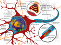 Complete neuron cell diagram
 -
 Medullated nerve fibers stained with silver nitrate


See also
References
- ^ "node of Ranvier". Lexico UK English Dictionary. Oxford University Press. Archived from the original on October 16, 2021.
- ^ "node of Ranvier". Merriam-Webster.com Dictionary.
- S2CID 6743084.
- ^ S2CID 6743084.
- S2CID 10252129.
- ^ Black, J.A., Sontheimer, H., Oh, Y., and Waxman, S.G. (1995). In The Axon, S. Waxman, J. Kocsis, and P. Stys, eds. Oxford University Press, New York, pp. 116–143.
- PMID 13449102.
- S2CID 7168889.
- PMID 9278538.
- S2CID 57536809.
- PMID 22219296.
- ^ PMID 17460780.
- S2CID 14537696.
- S2CID 17410200.
- PMID 20980605.
- Who Named It?
- S2CID 20120269.
- ^ Barbara J.G. (2005). "Les étranglements annulaires de Louis Ranvier (1871)" (PDF). Lettre des Neurosciences. 28: 3–5.
External links
- Cell Centered Database – Node of Ranvier
- Anatomy photo: nervous/pns/nerve2/nerve5 - Comparative Organology at University of California, Davis – "PNS, nerve (LM, Medium)"
| Authority control databases: National |
|---|