
Nuclear medicine
| Nuclear medicine | |
|---|---|
PET scan | |
| ICD-10-PCS | C |
| ICD-9 | 92 |
| MeSH | D009683 |
| OPS-301 code | 3-70-3-72, 8-53 |
Nuclear medicine or nucleology
Diagnostic medical imaging
Nuclear medicine, also known as nucleology, is a medical specialty that uses radioactive substances to diagnose and treat diseases. Unlike traditional radiology, which records radiation transmitted through the body from external sources like X-ray generators, nuclear imaging records radiation emitted from within the body. The focus of nuclear medicine scans is on the function of organs and tissues rather than their anatomy. This is why it is referred to as a physiological imaging modality. Nuclear medicine utilises two primary imaging modalities, single photon emission computed tomography (SPECT) and positron emission tomography (PET) scans. These are commonly used in the field. It is important to note that SPECT and PET scans are the two most common imaging modalities in nuclear medicine.
Diagnostic
In nuclear medicine imaging,
There are several techniques of diagnostic nuclear medicine.
- 2D: Scintigraphy ("scint") is the use of internal radionuclides to create two-dimensional images.[3]
-
 A nuclear medicine whole body bone scan. The nuclear medicine whole body bone scan is generally used in evaluations of various bone-related pathology, such as for bone pain, stress fracture, nonmalignant bone lesions, bone infections, or the spread of cancer to the bone.
 -
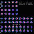 Nuclear medicine myocardial perfusion scan with thallium-201 for the rest images (bottom rows) and Tc-Sestamibi for the stress images (top rows). The nuclear medicine myocardial perfusion scan plays a pivotal role in the non-invasive evaluation of coronary artery disease. The study not only identifies patients with coronary artery disease; it also provides overall prognostic information or overall risk of adverse cardiac events for the patient.
 -
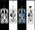
 A nuclear medicine parathyroid scan demonstrates a parathyroid adenoma adjacent to the left inferior pole of the thyroid gland. The above study was performed with Technetium-Sestamibi (1st column) and iodine-123 (2nd column) simultaneous imaging and the subtraction technique (3rd column).
 -
 Normal hepatobiliary scan (HIDA scan). The nuclear medicine hepatobiliary scan is clinically useful in the detection of the gallbladder disease.
 -
 Normal pulmonary ventilation and perfusion (V/Q) scan. The nuclear medicine V/Q scan is useful in the evaluation of pulmonary embolism.
 -
 Thyroid scan with iodine-123 for evaluation of hyperthyroidism.


- 3D: SPECT is a 3D tomographic technique that uses gamma camera data from many projections and can be reconstructed in different planes. Positron emission tomography(PET) uses coincidence detection to image functional processes.
-
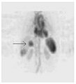 A nuclear medicine SPECT liver scan with technetium-99m labeled autologous red blood cells. A focus of high uptake (arrow) in the liver is consistent with a hemangioma.
 -
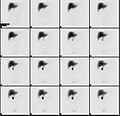
 Maximum intensity projection (MIP) of a whole-body positron emission tomography (PET) acquisition of a 79 kg female after intravenous injection of 371 MBq of 18F-FDG (one hour prior measurement).


Nuclear medicine tests differ from most other imaging modalities in that nuclear medicine scans primarily show the physiological function of the system being investigated as opposed to traditional anatomical imaging such as CT or MRI. Nuclear medicine imaging studies are generally more organ-, tissue- or disease-specific (e.g.: lungs scan, heart scan, bone scan, brain scan, tumor, infection, Parkinson etc.) than those in conventional radiology imaging, which focus on a particular section of the body (e.g.: chest X-ray, abdomen/pelvis CT scan, head CT scan, etc.). In addition, there are nuclear medicine studies that allow imaging of the whole body based on certain cellular receptors or functions. Examples are whole body

While the ability of nuclear metabolism to image disease processes from differences in metabolism is unsurpassed, it is not unique. Certain techniques such as
Diagnostic tests in nuclear medicine exploit the way that the body handles substances differently when there is disease or pathology present. The radionuclide introduced into the body is often chemically bound to a complex that acts characteristically within the body; this is commonly known as a tracer. In the presence of disease, a tracer will often be distributed around the body and/or processed differently. For example, the ligand methylene-diphosphonate (MDP) can be preferentially taken up by bone. By chemically attaching technetium-99m to MDP, radioactivity can be transported and attached to bone via the hydroxyapatite for imaging. Any increased physiological function, such as due to a fracture in the bone, will usually mean increased concentration of the tracer. This often results in the appearance of a "hot spot", which is a focal increase in radio accumulation or a general increase in radio accumulation throughout the physiological system. Some disease processes result in the exclusion of a tracer, resulting in the appearance of a "cold spot". Many tracer complexes have been developed to image or treat many different organs, glands, and physiological processes.
Hybrid scanning techniques
In some centers, the nuclear medicine scans can be superimposed, using software or hybrid cameras, on images from modalities such as CT or MRI to highlight the part of the body in which the radiopharmaceutical is concentrated. This practice is often referred to as image fusion or co-registration, for example SPECT/CT and PET/CT. The fusion imaging technique in nuclear medicine provides information about the anatomy and function, which would otherwise be unavailable or would require a more invasive procedure or surgery.
-
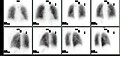
 Normal whole body PET/CT scan withFDG-18. The whole body PET/CT scan is commonly used in the detection, staging and follow-up of various cancers.
 -
 Abnormal whole body PET/CT scan with multiple metastases from a cancer. The whole body PET/CT scan has become an important tool in the evaluation of cancer.


Practical concerns in nuclear imaging
Although the risks of low-level radiation exposures are not well understood, a cautious approach has been universally adopted that all human radiation exposures should be kept As Low As Reasonably Practicable, "ALARP". (Originally, this was known as "As Low As Reasonably Achievable" (ALARA), but this has changed in modern draftings of the legislation to add more emphasis on the "Reasonably" and less on the "Achievable".)
Working with the ALARP principle, before a patient is exposed for a nuclear medicine examination, the benefit of the examination must be identified. This needs to take into account the particular circumstances of the patient in question, where appropriate. For instance, if a patient is unlikely to be able to tolerate a sufficient amount of the procedure to achieve a diagnosis, then it would be inappropriate to proceed with injecting the patient with the radioactive tracer.
When the benefit does justify the procedure, then the radiation exposure (the amount of radiation given to the patient) should also be kept as low as reasonably practicable. This means that the images produced in nuclear medicine should never be better than required for confident diagnosis. Giving larger radiation exposures can reduce the noise in an image and make it more photographically appealing, but if the clinical question can be answered without this level of detail, then this is inappropriate.
As a result, the radiation dose from nuclear medicine imaging varies greatly depending on the type of study. The effective radiation dose can be lower than or comparable to or can far exceed the general day-to-day environmental annual background radiation dose. Likewise, it can also be less than, in the range of, or higher than the radiation dose from an abdomen/pelvis CT scan.
Some nuclear medicine procedures require special patient preparation before the study to obtain the most accurate result. Pre-imaging preparations may include dietary preparation or the withholding of certain medications. Patients are encouraged to consult with the nuclear medicine department prior to a scan.
Analysis
The result of the nuclear medicine imaging process is a dataset comprising one or more images. In multi-image datasets the array of images may represent a time sequence (i.e. cine or movie) often called a "dynamic" dataset, a cardiac gated time sequence, or a spatial sequence where the gamma-camera is moved relative to the patient.
The nuclear medicine computer may require millions of lines of source code to provide quantitative analysis packages for each of the specific imaging techniques available in nuclear medicine.[citation needed]
Time sequences can be further analysed using
Interventional nuclear medicine
Radionuclide therapy can be used to treat conditions such as hyperthyroidism, thyroid cancer, skin cancer and blood disorders.
In nuclear medicine therapy, the radiation treatment dose is administered internally (e.g. intravenous or oral routes) or externally direct above the area to treat in form of a compound (e.g. in case of skin cancer).
The radiopharmaceuticals used in nuclear medicine therapy emit ionizing radiation that travels only a short distance, thereby minimizing unwanted side effects and damage to noninvolved organs or nearby structures. Most nuclear medicine therapies can be performed as outpatient procedures since there are few side effects from the treatment and the radiation exposure to the general public can be kept within a safe limit.
Common nuclear medicine
| Substance | Condition |
|---|---|
| Iodine-131-sodium iodide | hyperthyroidism and thyroid cancer |
| Yttrium-90-ibritumomab tiuxetan (Zevalin) and Iodine-131-tositumomab (Bexxar) | refractory lymphoma |
| 131I-MIBG ( metaiodobenzylguanidine )
|
neuroendocrine tumors |
Samarium-153 or Strontium-89
|
palliative bone pain treatment |
| Rhenium-188 | squamous cell carcinoma or basal cell carcinoma of the skin
|
In some centers the nuclear medicine department may also use implanted capsules of isotopes (brachytherapy) to treat cancer.
Commonly used radiation sources (radionuclides) for brachytherapy[4]
| Radionuclide | Type | Half-life | Energy |
|---|---|---|---|
| Caesium-137 (137Cs) | γ-ray | 30.17 years | 0.662 MeV |
| Cobalt-60 (60Co) | γ-ray | 5.26 years | 1.17, 1.33 MeV |
| Iridium-192 (192Ir) | β−-particles | 73.8 days | 0.38 MeV (mean) |
| Iodine-125 (125I) | γ-rays | 59.6 days | 27.4, 31.4 and 35.5 keV |
Palladium-103 (103Pd)
|
γ-ray | 17.0 days | 21 keV (mean) |
Ruthenium-106 (106Ru)
|
β−-particles | 1.02 years | 3.54 MeV |
History
The history of nuclear medicine contains contributions from scientists across different disciplines in physics, chemistry, engineering, and medicine. The multidisciplinary nature of nuclear medicine makes it difficult for medical historians to determine the birthdate of nuclear medicine. This can probably be best placed between the discovery of artificial radioactivity in 1934 and the production of radionuclides by Oak Ridge National Laboratory for medicine-related use, in 1946.[5]
The origins of this medical idea date back as far as the mid-1920s in
Many historians consider the discovery of artificially produced radionuclides by
Nuclear medicine gained public recognition as a potential specialty when on May 11, 1946, an article in the Journal of the American Medical Association (JAMA) by Massachusetts General Hospital's Dr.
Widespread clinical use of nuclear medicine began in the early 1950s, as knowledge expanded about radionuclides, detection of radioactivity, and using certain radionuclides to trace biochemical processes. Pioneering works by
By the early 1960s, in southern
By the 1970s most organs of the body could be visualized using nuclear medicine procedures. In 1971, American Medical Association officially recognized nuclear medicine as a medical specialty.[16] In 1972, the American Board of Nuclear Medicine was established, and in 1974, the American Osteopathic Board of Nuclear Medicine was established, cementing nuclear medicine as a stand-alone medical specialty.
In the 1980s, radiopharmaceuticals were designed for use in diagnosis of heart disease. The development of single photon emission computed tomography (SPECT), around the same time, led to three-dimensional reconstruction of the heart and establishment of the field of nuclear cardiology.
More recent developments in nuclear medicine include the invention of the first positron emission tomography scanner (PET). The concept of emission and transmission tomography, later developed into single photon emission computed tomography (SPECT), was introduced by David E. Kuhl and Roy Edwards in the late 1950s.[citation needed] Their work led to the design and construction of several tomographic instruments at the University of Pennsylvania. Tomographic imaging techniques were further developed at the Washington University School of Medicine. These innovations led to fusion imaging with SPECT and CT by Bruce Hasegawa from University of California, San Francisco (UCSF), and the first PET/CT prototype by D. W. Townsend from University of Pittsburgh in 1998.[citation needed]
PET and PET/CT imaging experienced slower growth in its early years owing to the cost of the modality and the requirement for an on-site or nearby cyclotron. However, an administrative decision to approve medical reimbursement of limited PET and PET/CT applications in oncology has led to phenomenal growth and widespread acceptance over the last few years, which also was facilitated by establishing 18F-labelled tracers for standard procedures, allowing work at non-cyclotron-equipped sites. PET/CT imaging is now an integral part of oncology for diagnosis, staging and treatment monitoring. A fully integrated MRI/PET scanner is on the market from early 2011.[citation needed]
Sources of radionuclides
99mTc is normally supplied to hospitals through a
The most commonly used radioisotope in PET,
| isotope | symbol | Z | T1/2 | decay | gamma (keV) | Maximum β
energy (keV) / Abundance[21] |
|---|---|---|---|---|---|---|
| Imaging: | ||||||
| fluorine-18 | 18F | 9 | 109.77 m | β+ | 511 (193%) | 634 (97%) |
gallium-67 |
67Ga | 31 | 3.26 d | ec | 93 (39%), 185 (21%), 300 (17%) |
- |
krypton-81m |
81mKr | 36 | 13.1 s | IT | 190 (68%) | - |
| rubidium-82 | 82Rb | 37 | 1.27 m | β+ | 511 (191%) | 3381 (81.8%)
2605 (13.1%) 1906 (0.14%) 1209 (0.32%) |
| nitrogen-13 | 13N | 7 | 9.97 m | β+ | 511 (200%) | 1198 (99.8%) |
| technetium-99m | 99mTc | 43 | 6.01 h | IT | 140 (89%) | - |
| indium-111 | 111In | 49 | 2.80 d | ec | 171 (90%), 245 (94%) |
- |
| iodine-123 | 123I | 53 | 13.3 h | ec | 159 (83%) | - |
xenon-133 |
133Xe | 54 | 5.24 d | β− | 81 (31%) | 346 (99.1%)
267 (0.9%) |
thallium-201 |
201Tl | 81 | 3.04 d | ec | 69–83* (94%), 167 (10%) |
- |
| Therapy: | ||||||
| yttrium-90 | 90Y | 39 | 2.67 d | β− | - | 2279 (99.98%) |
| iodine-131 | 131I | 53 | 8.02 d | β− | 364 (81%) | 807 (0.4%)
606 (89.4%) 334 (7.2%) 248 (2.1%) |
| lutetium-177 | 177Lu | 71 | 6.65 d | β− | 113 (6.6%),
208 (11%) |
498 (79.3%)
385 (9.1%) 177 (11.6%) |
|
Z = atomic number, the number of protons; T1/2 = half-life; decay = mode of decay | ||||||
A typical nuclear medicine study involves administration of a
The most commonly used intravenous radionuclides are technetium-99m, iodine-123, iodine-131, thallium-201, gallium-67, fluorine-18
Policies and procedures
Radiation dose
A patient undergoing a nuclear medicine procedure will receive a radiation dose. Under present international guidelines it is assumed that any radiation dose, however small, presents a risk. The radiation dose delivered to a patient in a nuclear medicine investigation, though unproven, is generally accepted to present a very small risk of inducing cancer. In this respect it is similar to the risk from X-ray investigations except that the dose is delivered internally rather than from an external source such as an X-ray machine, and dosage amounts are typically significantly higher than those of X-rays.
The radiation dose from a nuclear medicine investigation is expressed as an
Effective doses can range from 6 μSv (0.006 mSv) for a 3 MBq chromium-51 EDTA measurement of glomerular filtration rate to 11.2 mSv (11,200 μSv) for an 80 MBq thallium-201 myocardial imaging procedure. The common bone scan with 600 MBq of technetium-99m MDP has an effective dose of approximately 2.9 mSv (2,900 μSv).[23]
Formerly, units of measurement were the
Regulatory frameworks and guidelines
Different countries around the world maintain regulatory frameworks that are responsible for the management and use of radionuclides in different medical settings. For example, in the US, the Nuclear Regulatory Commission (NRC) and the Food and Drug Administration (FDA) have guidelines in place for hospitals to follow.[25] With the NRC, if radioactive materials aren't involved, like X-rays for example, they are not regulated by the agency and instead are regulated by the individual states.[26] International organizations, such as the International Atomic Energy Agency (IAEA), have regularly published different articles and guidelines for best practices in nuclear medicine as well as reporting on emerging technologies in nuclear medicine.[27][28] Other factors that are considered in nuclear medicine include a patient's medical history as well as post-treatment management. Groups like International Commission on Radiological Protection have published information on how to manage the release of patients from a hospital with unsealed radionuclides.[29]
See also
- Human subject research
- List of Nuclear Medicine Societies
- Nuclear medicine physician
- Nuclear pharmacy
- Nuclear technology
- Radiographer
References
- ^ What is nucleology?
- ^ "Nuclear Medicine". Archived from the original on 27 February 2015. Retrieved 20 August 2015.
- ^ scintigraphy Citing: Dorland's Medical Dictionary for Health Consumers, 2007 by Saunders; Saunders Comprehensive Veterinary Dictionary, 3 ed. 2007; McGraw-Hill Concise Dictionary of Modern Medicine, 2002 by The McGraw-Hill Companies
- ^ "Nuclear Wallet Cards". Retrieved 20 August 2015.
- ^ PMID 388628.
- ^ Donner Laboratory: The Birthplace of. Nuclear Medicine
- ^ "Important Moments in the History of Nuclear Medicine". Archived from the original on 2013-12-14. Retrieved 2012-01-03.
- PMID 21025609.
- PMID 20274882.
- ISBN 978-0-8016-7701-4.
- .
- PMID 4140398.
- PMID 705327.
- PMID 7351547.
- ^ Roland PE [in Swedish], Friberg L [in Swedish] (1985). "Localization of cortical areas activated by thinking". Journal of Neurophysiology. Vol. 53, no. 5. pp. 1219–1243.
- ^ "What is nuclear medicine" (PDF). Society of Nuclear Medicine. Archived from the original (PDF) on 2016-01-17. Retrieved 2009-01-17.
- ^ "Canada permanently closes NRU research reactor". Nuclear Engineering International. 6 April 2018.
- ISBN 978-0-932004-80-2
- ^ Table of Radioactive Isotopes Archived 2004-12-04 at the Wayback Machine
- PMID 26085854.
- ^ "Données atomiques et nucléaires". Laboratoire National Henri Becquerel. Retrieved 24 October 2022.
- ^ Technegas a radioaerosol invented in Australia by Dr Bill Burch and Dr Richard Fawdry
- ^ Administration of Radioactive Substances Advisory Committee (19 February 2021). "ARSAC notes for guidance" (pdf). GOV.UK. Public Health England.
- ^ Chandler, David (2011-03-28). "Explained: rad, rem, sieverts, becquerels". MIT News | Massachusetts Institute of Technology. Retrieved 2021-04-25.
- S2CID 230547683, retrieved 2021-04-25
- ^ "Nuclear Medicine: What It Is – and Isn't". NRC Web. 2020-06-08. Retrieved 2021-04-25.
- ^ "IAEA Safety Standards and medical exposure". www.iaea.org. 2017-10-30. Retrieved 2021-04-25.
- ^ "Human Health Campus – Nuclear Medicine". humanhealth.iaea.org. 21 February 2020. Retrieved 2021-04-25.
- S2CID 43014655.
Further reading
- Mas JC (2008). A Patient's Guide to Nuclear Medicine Procedures: English-Spanish. Society of Nuclear Medicine. ISBN 978-0-9726478-9-2.
- Taylor A, Schuster DM, Naomi Alazraki N (2000). A Clinicians' Guide to Nuclear Medicine (2nd ed.). Society of Nuclear Medicine. ISBN 978-0-932004-72-7.
- Shumate MJ, Kooby DA, Alazraki NP (2007). A Clinician's Guide to Nuclear Oncology: Practical Molecular Imaging and Radionuclide Therapies. Society of Nuclear Medicine. ISBN 978-0-9726478-8-5.
- Ell P, Gambhir S (2004). Nuclear Medicine in Clinical Diagnosis and Treatment. Churchill Livingstone. p. 1950. ISBN 978-0-443-07312-0.
- Jones DW, Hogg P, Seeram E (2013). Practical SPECT/CT In Nuclear Medicine. Springer. ISBN 978-1447147022.
External links
- Nuclear medicine at Curlie
- Solving the Medical Isotope Crisis Hearing before the Subcommittee on Energy and Environment of the Committee on Energy and Commerce, House of Representatives, One Hundred Eleventh Congress, First Session, September 9, 2009
| |||||||||||||||||||||||||||||||
| |||||||||||||||||||||||||||||||
| Gas |
| ||||||||||||||||||||||||||||||
Molten-salt |
| ||||||||||||||||||||||||||||||
(fast-neutron)
| Generation IV | |
|---|---|
confinement | |
|---|---|
| Magnetic | |
| Inertial | |
| Other | |