
Onychophora
| Onychophora Temporal range:
| |
|---|---|

| |
| Oroperipatus sp. (Peripatidae) | |

| |
| Ooperipatellus sp. (Peripatopsidae) | |
| Scientific classification | |
| Domain: | Eukaryota |
| Kingdom: | Animalia |
| Subkingdom: | Eumetazoa |
| Clade: | ParaHoxozoa |
| Clade: | Bilateria |
| Clade: | Nephrozoa |
| (unranked): | Protostomia |
| Superphylum: | Ecdysozoa |
| (unranked): | Panarthropoda |
| Phylum: | Onychophora Grube, 1850 |
| Subgroups | |
| |

| |
| Global range of Onychophora extant Peripatidae fossils
| |
Onychophora
Anatomy
Velvet worms are
Appendages
The stub feet that characterise the velvet worms are
- Crural glands are situated at the shoulder of the legs, extending into the body cavity. They open outwards at the crural papillae—small wart-like bumps on the belly side of the leg—and secrete chemical messenger materials called pheromones. Their name comes from the Latin cruralis meaning "of the legs".[13]
- Coxal vesicles are pouches located on the belly side of the leg, which can be everted and probably serve in water absorption. They belong to the family Peripatidae and are named from coxa, the Latin word for "hip".[14]
On each foot is a pair of retractable, hardened (sclerotised)
- On the first head segment is a pair of slender blind species. In front of these, in many Australian species, are various dimples, the function of which is not yet clear. It appears that in at least some species, these serve in the transfer of sperm-cell packages (spermatophores).[citation needed]
- On the belly side of the second head segment is the labrum, a mouth opening surrounded by sensitive "lips". In the velvet worms, this structure is a muscular outgrowth of the throat, so, despite its name, it is probably not homologous to the labrum of the Arthropoda and is used for feeding. Deep within the oral cavity lie the sharp, crescent-shaped "jaws", or mandibles, which are strongly hardened and resemble the claws of the feet, with which they are serially homologous;[17] early in development, the jaw appendages have a position and shape similar to the subsequent legs.[18] The jaws are divided into internal and external mandibles and their concave surface bears fine denticles. They move backward and forward in a longitudinal direction, tearing apart the prey, apparently moved in one direction by musculature and the other by hydrostatic pressure.[17] The claws are made of sclerotised α-chitin, reinforced with phenols and quinones, and have a uniform composition – except that there is a higher concentration of calcium towards the tip, presumably affording greater strength.[17]
The surface of the mandibles is smooth, with no ornamentation.[19] The cuticle in the mandibles (and claws) is distinct from the rest of the body. It has an inner and outer component; the outer component has just two layers (whereas body cuticle has four), and these outer layers (in particular the inner epicuticle) are dehydrated and strongly tanned, affording toughness.[19]
Slime glands

On the third head segment, to the left and right of the mouth, are two openings designated "oral papillae". Within these are a pair of large, heavily internally branched slime glands. These lie roughly in the centre of the body and secrete a sort of milky-white slime, which is used to ensnare
Skin and muscle
Unlike the arthropods, velvet worms do not possess a rigid
Within the connective tissue lie three continuous layers of unspecialised smooth muscular tissue. The relatively thick outer layer is composed of annular muscles, and the similarly voluminous inner layer of longitudinal muscles. Between them lie thin diagonal muscles that wind backward and forward along the body axis in a spiral. Between the annular and diagonal muscles exist fine blood vessels, which lie below the superficially recognisable transverse rings of the skin and are responsible for the pseudo-segmented markings.[12] Beneath the internal muscle layer lies the body cavity. In cross-section, this is divided into three regions by so-called dorso-ventral muscles, which run from the middle of the underbelly through to the edges of the upper side: a central midsection and on the left and right, two side regions that also include the legs.[citation needed]
Circulation
The body cavity is known as a "pseudocoel", or
The haemocoel is divided by a horizontal partition, the diaphragm, into two parts: The pericardial sinus along the back and the perivisceral sinus along the belly. The former encloses the tube-like heart, and the latter, the other organs. The diaphragm is perforated in many places, enabling the exchange of fluids between the two cavities.[citation needed] The heart itself is a tube of annular muscles consisting of epithelial tissues, with two lateral openings (ostia) per segment. While it is not known whether the rear end is open or closed, from the front, it opens directly into the body cavity.
Since there are no blood vessels, apart from the fine vessels running between the muscle layers of the body wall and a pair of arteries that supply the antennae, this is referred to as an
Respiration
Oxygen uptake occurs to an extent via simple
The walls of these structures, which are less than three micrometers thick in their entirety, consist only of an extremely thin membrane through which oxygen can easily diffuse. The tracheae originate at tiny openings, the spiracles, which themselves are clustered together in dent-like recesses of the outer skin, the atria. The number of "tracheae bundles" thus formed is on average around 75 bundles per body segment; they accumulate most densely on the back of the organism.[citation needed]
Unlike the arthropods, the velvet worms are unable to control the openings of their tracheae; the tracheae are always open, entailing considerable water loss in
Oxygen transport is helped by the oxygen carrier hemocyanin.[28]
Digestion

The digestive tract begins slightly behind the head, the mouth lying on the underside a little way from the frontmost point of the body. Here, prey can be mechanically dismembered by the mandibles with their covering of fine toothlets. Two
The throat itself is very muscular, serving to absorb the partially liquified food and to pump it, via the
On entering the central intestine, food particles are coated with a mucus-based
In almost every segment is a pair of excretory organs called nephridia, which are derived from coelom tissue. Each consists of a small pouch that is connected, via a flagellated conductor called a nephridioduct, to an opening at the base of the nearest leg known as a nephridiopore. The pouch is occupied by special cells called podocytes, which facilitate ultrafiltration of the blood through the partition between haemocoelom and nephridium.
The composition of the
A pair of former nephridia in the head were converted secondarily into the salivary glands, while another pair in the final segment of male specimens now serve as glands that apparently play a role in reproduction.[29]
Sensation
The entire body, including the stub feet, is littered with numerous papillae: warty protrusions responsive to touch that carry a
Except in a few (typically
The
Reproduction
Both sexes possess pairs of gonads, opening via a channel called a gonoduct into a common genital opening, the gonopore, which is located on the rear ventral side. Both the gonads and the gonoduct are derived from true coelom tissue.[citation needed]

In females, the two
Males possess two separate
There are different mating procedures: In some species males deposit their spermatophore directly into the female's genitals opening, while others deposit it on the female's body, where the cuticle will collapse and allowing the sperm cells to migrate into the female. There are also Australian species where the male place their spermatophore on top of their head, which is then pressed against the female's genitals. In these species the head have elaborate structures like spikes, spines, hollow stylets, pits, and depressions, whose purpose is to either hold the sperm and / or assist in the sperm transfer to the female. The males of most species also secrete a pheromone from glands on the underside of the legs to attract females.[33]
Distribution and habitat
Distribution
Velvet worms live in all
Fossils have been found in Baltic amber, indicating that they were formerly more widespread in the Northern Hemisphere when conditions were more suitable.[37]
Habitat
Velvet worms always sparsely occupy the habitats where they are found: They are rare among the fauna which they are a part of.[citation needed]
All extant velvet worms are terrestrial (land-living) and prefer dark environments with high air humidity. They are found particularly in the
Some species of velvet worms are able to occupy human-modified land-uses, such as
Velvet worms are photophobic: They are repelled by bright light sources.[39] Because the danger of desiccation is greatest during the day and in dry weather, it is not surprising that velvet worms are usually most active at night and during rainy weather. Under cold or dry conditions, they actively seek out crevices in which they shift their body into a resting state.[40]
Slime

The Onychophora forcefully
The slime glands themselves are deep inside the body cavity, each at the end of a tube more than half the length of the body. The tube both conducts the fluid and stores it until it is required. The distance that the animal can propel the slime varies; usually it squirts it about a centimetre,[45] but the maximal range has variously been reported to be ten centimetres,[46] or even nearly a foot,[47] although accuracy drops with range.[48] It is not clear to what extent the range varies with the species and other factors. One squirt usually suffices to snare a prey item, although larger prey may be further immobilised by smaller squirts targeted at the limbs; additionally, the fangs of spiders are sometimes targeted.[48] Upon ejection, it forms a net of threads about twenty microns in diameter, with evenly spaced droplets of viscous adhesive fluid along their length.[45] It subsequently dries, shrinking, losing its stickiness, and becoming brittle.[45] Onychophora eat their dried slime when they can, which is appropriate, because it takes an onychophoran about 24 days to replenish an exhausted slime repository.[48]
The slime can account for up to 11% of the organism's dry weight
Behaviour
Locomotion

Velvet worms/Onychophora move in a slow and gradual motion that makes them difficult for prey to notice.[48] Their trunk is raised relatively high above the ground, and they walk with non-overlapping steps.[50] To move from place to place, the velvet worm crawls forward using its legs; unlike in arthropods, both legs of a pair are moved simultaneously. The claws of the feet are used only on hard, rough terrain where a firm grip is needed; on soft substrates, such as moss, the velvet worm walks on the foot cushions at the base of the claws.[citation needed]
Actual locomotion is achieved less by the exertion of the leg muscles than by local changes of body length. This can be controlled using the annular and longitudinal muscles. If the annular muscles are contracted, the body cross-section is reduced, and the corresponding segment lengthens; this is the usual mode of operation of the hydrostatic skeleton as also employed by other worms. Due to the stretching, the legs of the segment concerned are lifted and swung forward. Local contraction of the longitudinal muscles then shortens the appropriate segment, and the legs, which are now in contact with the ground, are moved to the rear. This part of the locomotive cycle is the actual leg stroke that is responsible for forward movement. The individual stretches and contractions of the segments are coordinated by the nervous system such that contraction waves run the length of the body, each pair of legs swinging forward and then down and rearward in succession. Macroperipatus can reach speeds of up to four centimetres per second,[48] although speeds of around 6 body-lengths per minute are more typical.[51] The body gets longer and narrower as the animal picks up speed; the length of each leg also varies during each stride.[51]
Sociality
The brains of Onychophora, though small, are very complex; consequently, the organisms are capable of rather sophisticated social interactions.[52] Behaviour may vary from genus to genus, so this article reflects the most-studied genus, Euperipatoides.[52]
The Euperipatoides form social groups of up to fifteen individuals, usually closely related, which will typically live and hunt together. Groups usually live together; in drier regions an example of a shared home would be the moist interior of a rotting log. Group members are extremely aggressive towards individuals from other logs.[52] Dominance is achieved through aggression and maintained through submissive behaviour.[52] After a kill, the dominant female always feeds first, followed in turn by the other females, then males, then the young.[52]
When assessing other individuals, individuals often measure one another up by running their antennae down the length of the other individual.[52] Once hierarchy has been established, pairs of individuals will often cluster together to form an "aggregate"; this is fastest in male-female pairings, followed by pairs of females, then pairs of males.[52]
Social hierarchy is established by a number of interactions: Higher-ranking individuals will chase and bite their subordinates while the latter are trying to crawl on top of them.[52] Juveniles never engage in aggressive behaviour, but climb on top of adults, which tolerate their presence on their backs.[52] Hierarchy is quickly established among individuals from a single group, but not among organisms from different groups; these are substantially more aggressive and very rarely climb one another or form aggregates.[52] Individuals within an individual log are usually closely related; especially so with males. This may be related to the intense aggression between unrelated females.[52]
Feeding
.jpg)
Velvet worms are
The most energetically favourable prey are two-fifths the size of the hunting onychophoran.[48] Ninety percent of the time involved in eating prey is spent ingesting it; re-ingestion of the slime used to trap the insect is performed while the onychophoran locates a suitable place to puncture the prey, and this phase accounts for around 8% of the feeding time, with the remaining time evenly split between examining, squirting, and injecting the prey.[48] In some cases, chunks of the prey item are bitten off and swallowed; undigestable components take around 18 hours to pass through the digestive tract.[17]
Onychophora probably do not primarily use vision to detect their prey; although their tiny eyes do have a good image-forming capacity, their forward vision is obscured by their antennae;[48] their nocturnal habit also limits the utility of eyesight. Air currents, formed by prey motion, are thought to be the primary mode of locating prey; the role of scent, if any, is unclear.[48] Because it takes so long to ingest a prey item, hunting mainly happens around dusk; the onychophorans will abandon their prey at sunrise.[48] This predatory way of life is probably a consequence of the velvet worm's need to remain moist. Due to the continual risk of desiccation, often only a few hours per day are available for finding food. This leads to a strong selection for a low cost-benefit ratio, which cannot be achieved with a herbivorous diet.[citation needed]
Velvet worms literally creep up on their prey, with their smooth, gradual and fluid movement escaping detection.[48] Once they reach their prey, they touch it very softly with their antennae to assess its size and nutritional value. After each poke, the antenna is hastily retracted to avoid alerting the prey.[48] This investigation may last anywhere upwards of ten seconds, until the velvet worm makes a decision as to whether to attack it, or until it disturbs the prey and the prey flees.[48] Hungry Onychophora spend less time investigating their prey and are quicker to apply their slime.[48] Once slime has been squirted, Onychophora are determined to pursue and devour their prey, in order to recoup the energy investment. They have been observed to spend up to ten minutes searching for removed prey, after which they return to their slime to eat it.[48] In the case of smaller prey, they may opt not to use slime at all.[48] Subsequently, a soft part of the prey item (usually a joint membrane in arthropod prey) is identified, punctured with a bite from the jaws, and injected with saliva. This kills the prey very quickly and begins a slower process of digestion.[48] While the onychophoran waits for the prey to digest, it salivates on its slime and begins to eat it (and anything attached to it). It subsequently tugs and slices at the earlier perforation to allow access to the now-liquefied interior of its prey.[48] The jaws operate by moving backwards and forwards along the axis of the body (not in a side-to-side clipping motion as in arthropods), conceivably using a pairing of musculature and hydrostatic pressure.[17] The pharynx is specially adapted for sucking, to extract the liquefied tissue; the arrangement of the jaws about the tongue and lip papillae ensures a tight seal and the establishment of suction.[17] In social groups, the dominant female is the first to feed, not permitting competitors access to the prey item for the first hour of feeding. Subsequently, subordinate individuals begin to feed. The number of males reaches a peak after females start to leave the prey item.[52] After feeding, individuals clean their antennae and mouth parts before re-joining the rest of their group.[52]
Reproduction and life-cycle
Almost all species of velvet worm reproduce sexually. The sole exception is
The detailed process by which this is achieved is in most cases still unknown, a true
In a recent peer-reviewed paper published in the "Journal of Zoology," researchers discovered that certain species of Peripatus exhibit a unique form of parental care. Unlike most invertebrates, where parental involvement is minimal, female Peripatus were observed actively guarding their eggs and even providing protection to their offspring after hatching. This finding challenges the conventional understanding of reproductive behavior in invertebrates and highlights the diversity of parenting strategies in the animal kingdom.[55]
- Ovipary occurs solely in the Peripatopsidae, often in regions with erratic food supply or unsettled climate. In these cases, the Maternalcare is unknown.
- The majority of species are ovoviviparous: the medium-sized eggs, encased only by a double membrane, remain in the lecithotrophic. The young emerge from the eggs only a short time before birth. This probably represents the velvet worm's original mode of reproduction, i.e., both oviparous and viviparous species developed from ovoviviparous species.
- True live-bearing species are found in both families, particularly in tropical regions with a stable climate and regular food supply throughout the year. The embryos develop from eggs only micrometres in size and are nourished in the uterus by their mother, hence the description "matrotrophic". The supply of food takes place either via a secretion from the mother directly into the uterus or via a genuine tissue connection between the epithelium of the uterus and the developing embryo, known as a placenta. The former is found only outside the American continents, while the latter occurs primarily in America and the Caribbean and more rarely in the Old World. The gestation period can amount to up to 15 months, at the end of which the offspring emerge in an advanced stage of development. The embryos found in the uterus of a single female do not necessarily have to be of the same age; it is quite possible for there to be offspring at different stages of development and descended from different males. In some species, young tend to be released only at certain points in the year.[56]
A female can have between 1 and 23 offspring per year; development from fertilized ovum to adult takes between 6 and 17 months and does not have a larval stage. This is probably also the original mode of development. Velvet worms have been known to live for up to six years.
Ecology
The velvet worm's important predators are primarily various spiders and centipedes, along with
The South African species
Conservation
The global
- Mesoperipatus tholloni(Data Deficient)
- Plicatoperipatus jamaicensis(Near Threatened)
- Peripatoides indigo (Vulnerable)
- Peripatoides suteri (Vulnerable)
- Peripatopsis alba (Vulnerable)
- Peripatopsis clavigera (Vulnerable)
- Macroperipatus insularis (Endangered)
- Leucopatus anophthalmus(Endangered)
- Opisthopatus roseus (Critically Endangered)
- Peripatopsis leonina (Critically Endangered)
- Speleoperipatus spelaeus (Critically Endangered)[59]
The primary threat comes from destruction and fragmentation of velvet worm habitat due to industrialisation, draining of wetlands, and slash-and-burn agriculture. Many species also have naturally low population densities and closely restricted geographic ranges; as a result, relatively small localised disturbances of important ecosystems can lead to the extinction of entire populations or species. Collection of specimens for universities or research institutes also plays a role on a local scale.[60] There is a very pronounced difference in the protection afforded to velvet worms between regions: in some countries, such as South Africa, there are restrictions on both collecting and exporting, while in others, such as Australia, only export restrictions exist. Many countries offer no specific safeguards at all. Tasmania has a protection programme that is unique worldwide: one region of forest has its own velvet worm conservation plan, which is tailored to a particular velvet worm species.[60]
Phylogeny

In their present forms, the velvet worms are probably very closely related to the arthropods, a very extensive taxon that incorporates, for instance, the crustaceans, insects, and arachnids. They share, among other things, an exoskeleton consisting of α-chitin and non-collagenous proteins; gonads and waste-elimination organs enclosed in true coelom tissue; an open blood system with a tubular heart situated at the rear; an abdominal cavity divided into pericardial and perivisceral cavities; respiration via tracheae; and similar embryonic development. Segmentation, with two body appendages per segment, is also a shared feature.
However, the antennae, mandibles, and oral papillae of velvet worms are probably not homologous to the corresponding features in arthropods; i.e., they probably developed independently.
Another closely related group are the comparatively obscure water bears (
Together, the velvet worms, arthropods, and water bears form a
| Panarthropoda |
| ||||||||||||
.png)

For a long time, velvet worms were also considered related to the annelids. They share, among other things, a worm-like body; a thin and flexible outer skin; a layered musculature; paired waste-elimination organs; as well as a simply constructed brain and simple eyes. Decisive, however, was the existence of segmentation in both groups, with the segments showing only minor specialisation. The parapodia appendages found in annelids therefore correspond to the stump feet of the velvet worms. Within the Articulata hypothesis developed by Georges Cuvier, the velvet worms therefore formed an evolutionary link between the annelids and the arthropods: worm-like precursors first developed parapodia, which then developed further into stub feet as an intermediate link in the ultimate development of the arthropods' appendages. Due to their structural conservatism, the velvet worms were thus considered "living fossils". This perspective was expressed paradigmatically in the statement by the French zoologist A. Vandel:
- Onychophorans can be considered highly evolved annelids, adapted to terrestrial life, which announced prophetically the Arthropoda. They are a lateral branch which has endured from ancient times until today, without important modifications.
Modern taxonomy does not study criteria such as "higher" and "lower" states of development or distinctions between "main" and "side" branches—only family relationships indicated by
| Protostomia
|
| |||||||||||||||


Particularly characteristic of the Cycloneuralia is a ring of "circumoral" nerves around the mouth opening, which the proponents of the Ecdysozoa hypothesis also recognise in modified form in the details of the nerve patterns of the Panarthropoda. Both groups also share a common skin-shedding mechanism (
The "stem-group arthropod" hypothesis is very widely accepted, but some trees suggest that the onychophorans may occupy a different position; their brain anatomy is more closely related to that of the
- The Peripatopsidae exhibit relatively many characteristics that are perceived as original or "primitive". The number of leg pairs in this family range from 13 (in Ooperipatellus nanus[10]) to 29 (in Paraperipatus papuensis[65][66]).[67] Behind or between the last leg pair is the genital opening (gonopore).[67][68] Both oviparous and ovoviviparous, as well as genuinely viviparous, species exist, although the peripatopsids essentially lack a placenta. Their distribution is circumaustral, encompassing Australasia, South Africa, and Chile.[64]
- The Peripatidae exhibit a range of derivative features. They are longer, on average, than the Peripatopsidae and also have more legs. The number of leg pairs in this family range from 19 (in Plicatoperipatus jamaicensis[10]).[67][68] The gonopore is always between the penultimate leg pair.[67] None of the peripatid species are oviparous, and the overwhelming majority are viviparous. The females of many viviparous species develop a placenta with which to provide the growing embryo with nutrients. Distribution of the peripatids is restricted to the tropical and subtropical zones; in particular, they inhabit Central America, northern South America, Gabon, Northeast India, and Southeast Asia.[64]
Evolution

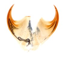
Certain fossils from the early Cambrian bear a striking resemblance to the velvet worms. These fossils, known collectively as the
.jpg)
Historically, all fossil Onychophora and lobopodians were lumped into the taxon
The low
The vagaries of the preservation process can make fossils difficult to interpret. Experiments on the decay and compaction of onychophora demonstrate difficulties in interpreting fossils; certain parts of living onychophora are visible only in certain conditions:
- The mouth may or may not be preserved;
- The claws may be re-oriented or lost;
- The leg width may increase or decrease; and
- The mud may be mistaken for organs.[80]
More significantly, features seen in fossils may be artefacts of the preservation process: For instance, "shoulder pads" may simply be the second row of legs coaxially compressed onto the body; branching "antennae" may in fact be produced through decay.[80]
References
- ISBN 978-0-7993-4689-3.[page needed]
- ISBN 978-0-231-13962-5.
- ISBN 978-0-03-025982-1.
Because they resemble worms with legs ... Superficially they resemble caterpillars, but have also been compared with slugs
- ISBN 978-0-313-33922-6.
- ^ PMID 27708504.
- ^ Fishelson, L. (1978). Zoology. Vol. 1 (3rd ed.). Israel: Hakibutz Hameuchad Publishing. p. 430.[verification needed]
- ^ doi:10.1071/IS18007.
- S2CID 55395265.
- PMID 21246983.
- ^ PMID 26124122– via ResearchGate.
- PMID 25788603.
- ^ ISBN 9780471042907.
- ^ "cruralis/crurale, cruralis M". Latin is Simple. Peter Waldert. Retrieved 2021-02-21.
- ^ "Cox, coxae, [f]". Latin is Simple. Peter Waldert. Retrieved 2021-02-21.
- ^ (PDF) from the original on 2022-10-10.
- S2CID 6755763.
- ^ PMID 25829018.
- S2CID 42865895.
- ^ PMID 18620280.
- arXiv:1511.00983. Archived from the originalon 2015-04-02.
- PMID 25780995.
- ISBN 978-90-04-08972-3.
- S2CID 83993887.
- ^ .
- JSTOR 92441.
- ^ "Velvet worm". The Australian Museum. January 18, 2022. Retrieved February 17, 2024.
- ^ "What is a velvet worm?". BBC Science Focus Magazine. July 5, 2022. Retrieved February 17, 2024.
- ^ Sizing Up the Onychophoran Genome: Repeats, Introns, and Gene Family Expansion Contribute to Genome Gigantism in Epiperipatus broadwayi
- ^ Holt, Jack (2010-02-07). "Introduction to the Onychophora". Protostomes > Ecdysozoa > Onychophora. Biol. 202: Systematic Biology. Selinsgrove, PA: Susquehanna University. Archived from the original on Oct 5, 2022. Retrieved 2022-11-01.
Excretory system: Pared metanephridia opening by their own ducts at each internal "segment". Anterior nephridia modified to salivary glands; posterior nephridia modified to gonopores.
- ^ PMID 18089073.
- ISBN 978-91-554-5613-9.[page needed]
- PMID 23284667.
- ^ "Velvet worm". The Australian Museum.
- ^ Kemp, S. (1913). "Preliminary note on a new genus of Onychophora from the N. E. Frontier of India". Records of the Indian Museum. 9: 241–242.
- S2CID 88237018.
- PMID 23284667.
- S2CID 85373762.
- .
- ^ "Velvet worm". The Australian Museum. Retrieved 2024-02-21.
- ^ Bang to Eternity and Betwixt: Cosmos. John Hussey. 2014-07-31.
- .
- S2CID 701697.
- ^ PMID 25780995.
- arXiv:1511.00983.
- ^ .
- ISBN 978-0-7993-4689-3.[page needed]
- ^ Harmer, Sidney Frederic; Shipley, Arthur Everett; et al. (1922). Peripatus, Myriapods, Insects. The Cambridge Natural Nistory. Vol. 5. Macmillan Company.[page needed]
- ^ S2CID 83545214.
- JSTOR 109116.
- ISBN 978-3-211-22134-1.
- ^ .
- ^ .
- ^ S2CID 86820446.
- .
- ^ Smith, J., & Jones, M. (2022). Maternal care in Peripatus: evidence for a hitherto unknown reproductive strategy in onychophorans. Journal of Zoology, 301 (2), 125–134.
- .
- ^ Mongenajera, J.; Barrientos, Z.; Aguilar, F. (1993). "Behaviour of Epiperipatus-biolleyi (Onychophora, Peripatidae) laboratory conditions". Revista de Biologica Tropical. 41 (3A): 689–696.
- .
- ^ "The IUCN Red List of Threatened Species". IUCN Red List of Threatened Species.
- ^ .
- PMID 17934206.
- PMID 16822744.
- ISBN 978-1-86977-849-1. Archived (PDF) from the original on 2022-10-10.)
{{cite book}}:|journal=ignored (help - ^ PMID 22930648.
- ISSN 2215-2075.
- S2CID 45711430.
- ^ .
- ^ ISSN 1175-5334– via ResearchGate.
- S2CID 88237018– via Biodiversity Heritage Library.
- ^ S2CID 205239797.
- S2CID 37935884.
- .
- S2CID 4313285.
- .
- JSTOR 1304204.
- ^ S2CID 53645124.
- .
- ^ JSTOR 3227105.
- PMID 34427671.
- ^ PMID 12947596.
External links
- Flickr
- I.S. Oliveira; L. Hering; G. Mayer. "Onychophora Website". Archived from the original on 14 June 2017.
- Youtube, The Slimy, Deadly Velvet Worm, Smithsonian Channel
- . Encyclopædia Britannica (11th ed.). 1911.
- Peripatus discussed on RNZ Critter of the Week, 13 Nov 2015