
Orbit (anatomy)
| Orbit | |
|---|---|
inferior rectus muscles; supraorbital foramen shown above the eye, and inferior orbital fissure inferolaterally. | |
| Details | |
| Identifiers | |
| Latin | orbita |
| MeSH | D009915 |
| TA98 | A02.1.00.067 |
| TA2 | 469 |
| FMA | 53074 |
| Anatomical terminology] | |
In
Structure
The orbits are conical or four-sided pyramidal cavities, which open into the midline of the face and point back into the head. Each consists of a base, an apex and four walls.[4]
Openings
There are two important
The
Bony walls
The bony walls of the orbital canal in humans do not derive from a single bone, but a mosaic of seven
The roof (superior wall) is formed primarily by the orbital plate frontal bone, and also the lesser wing of sphenoid near the apex of the orbit. The orbital surface presents medially by trochlear fovea and laterally by lacrimal fossa.[7]
The floor (inferior wall) is formed by the orbital surface of maxilla, the orbital surface of zygomatic bone and the minute orbital process of palatine bone. Medially, near the orbital margin, is located the groove for nasolacrimal duct. Near the middle of the floor, located infraorbital groove, which leads to the infraorbital foramen. The floor is separated from the lateral wall by inferior orbital fissure, which connects the orbit to pterygopalatine and infratemporal fossa.
The medial wall is formed primarily by the orbital plate of
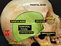
The lateral wall is formed by the frontal process of zygomatic and more posteriorly by the orbital plate of the greater wing of sphenoid. The bones meet at the zygomaticosphenoid suture. The lateral wall is the thickest wall of the orbit, important because it is the most exposed surface, highly vulnerable to blunt force trauma.
Borders
The base, orbital margin, which opens in the face, has four borders. The following bones take part in their formation:
- Superior margin: frontal bone
- Inferior margin: maxilla and zygomatic bone
- Medial margin: frontal bone and maxilla
- Lateral margin: zygomatic bone and frontal bone
Function
The orbit holds and protects the eyes.
Eye movement
The movement of the eye is controlled by six distinct extraocular muscles, a
Clinical significance
In the orbit, the surrounding fascia allows for smooth rotation and protects the orbital contents. If excessive tissue accumulates behind the ocular globe, the eye can protrude, or become exophthalmic.[4]

a. tear gland / lacrimal gland,
b. superior lacrimal punctum,
c. superior lacrimal canal,
d. tear sac / lacrimal sac,
e. inferior lacrimal punctum,
f. inferior lacrimal canal,
g. nasolacrimal canal
Enlargement of the lacrimal gland, located superotemporally within the orbit, produces protrusion of the eye inferiorly and medially (away from the location of the lacrimal gland). Lacrimal gland may be enlarged from inflammation (e.g. sarcoid) or neoplasm (e.g. lymphoma or adenoid cystic carcinoma).[9]
Tumors (e.g. glioma and meningioma of the optic nerve) within the cone formed by the horizontal rectus muscles produce axial protrusion (bulging forward) of the eye.
Additional images
This gallery of anatomic features needs cleanup to abide by the medical manual of style. ; please improve or remove the gallery accordingly. |
-
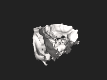
 Orbita
 -
 Medial wall of left orbit
 -
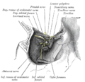 Dissection showing origins of right ocular muscles, and nerves entering by the superior orbital fissure
 -

-
 Lateral orbit nerves
 -
 Orbital cavity


References
- ^ "Orbit – Definition and More from the Free Merriam-Webster Dictionary". Retrieved 2010-03-26.
- ^ Orbit at the U.S. National Library of Medicine Medical Subject Headings (MeSH)
- ISBN 978-0-7817-6855-9.
- ^ Encyclopædia Britannica 2006 Ultimate Reference Suite DVD2009
- S2CID 43317074.
- PMID 25665284.
- ^ ISBN 978-07817-7525-0.
- S2CID 5567574.
- ISBN 9780721601878.
- PMID 12064074.
External links
- oph/2 at eMedicine – "Arterial Supply, Orbit"
- Anatomy photo:29:os-0501 at the SUNY Downstate Medical Center
- Atlas image: eye_5 at the University of Michigan Health System
- Atlas image: rsa2p4 at the University of Michigan Health System
- Interactive tutorial at anatome.ncl.ac.uk