
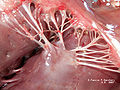
Papillary muscle
| Papillary muscle | |
|---|---|
Right ventricle. V.S. Ventricular septum. | |
| Details | |
| Identifiers | |
| Latin | musculus papillaris |
| MeSH | D010210 |
| TA98 | A12.1.00.022 |
| TA2 | 4043, 4066 |
| FMA | 12154 |
| Anatomical terms of muscle] | |
The papillary muscles are muscles located in the ventricles of the heart. They attach to the cusps of the
Structure
There are five total papillary muscles in the heart; three in the right ventricle and two in the left. The anterior, posterior, and septal papillary muscles of the right ventricle each attach via chordae tendineae to the tricuspid valve. The anterolateral and posteromedial papillary muscles of the left ventricle attach via chordae tendineae to the mitral valve.[2]
Blood supply
The mitral valve papillary muscles in the left ventricle are called the anterolateral and posteromedial muscles.[3]
- Anterolateral muscle blood supply: obtuse marginal branch(LCX)
- Posteromedial muscle blood supply: posterior interventricular artery(RCA)
The posteromedial muscle ruptures more frequently because it only has one source of blood supply, hence RCA occlusion can cause papillary muscle rupture.[3]
Function
The papillary muscles of both the right and left ventricles begin to contract shortly before
Clinical significance
Papillary muscle rupture can be caused by a
Additional images
-
 Opened chambers of the heart displaying papillary muscles and chordae tendineae
 -
 Papillary muscle infarction
 -
 Papillary muscles and chordae tendineae
 -
 Papillary muscles and chordae tendineae
 -
 Papillary muscles. Deep dissection.


See also
References
- ^ ISBN 978-0-7817-6274-8.
- ^ Netter's Atlas of Human Anatomy, plates 216B and 217A
- ^ PMID 21382906.
- .
- ISBN 978-0-7817-7153-5.
External links
- Anatomy photo:20:19-0106 at the SUNY Downstate Medical Center} — "Heart: The Right Atrioventricular (Tricupsid) Valve" (anterior, posterior, septal papillary muscles)
- Anatomy photo:20:26-0105 at the SUNY Downstate Medical Center — "Heart: The Left Atrioventricular (Mitral) Valve" (anterior, posterior papillary muscles)
- Atlas image: ht_rt_vent at the University of Michigan Health System} — "Right atrioventricular bundle branch, anterior view"
- Definition of Papillary muscle
- MedicineNet Search Results
| National | |
|---|---|
| Other | |