Pelger–Huët anomaly
| Pelger–Huët anomaly | |
|---|---|
| Other names | PHA[1] |
 | |
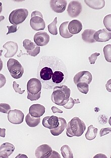
| blood smear of a patient with myelodysplastic syndrome: red blood cells showing marked poikilocytosis, in part related to post-splenectomy status, a central and hypogranular neutrophil with a pseudo-Pelger-Huet nucleus. | |
| Pronunciation |
|
| Specialty | Medical genetics |
Pelger–Huët anomaly is a blood laminopathy associated with the lamin B receptor,[2] wherein several types of white blood cells (neutrophils and eosinophils) have nuclei with unusual shape (being bilobed, peanut or dumbbell-shaped instead of the normal trilobed shape) and unusual structure (coarse and lumpy).[3]

It is a
Congenital Pelger–Huët anomaly
Is a benign
It is a genetic disorder with an autosomal dominant inheritance pattern.
Identifying Pelger–Huët anomaly is important to differentiate from bandemia with a left-shifted peripheral blood smear and neutrophilic band forms and from an increase in young neutrophilic forms that can be observed in association with infection.[citation needed]
Acquired or pseudo-Pelger–Huët anomaly
Anomalies resembling Pelger–Huët anomaly that are acquired rather than congenital have been described as pseudo Pelger–Huët anomaly. These can develop in the course of
In patients with these conditions, the pseudo–Pelger–Huët cells tend to appear late in the disease and often appear after considerable chemotherapy has been administered. The morphologic changes have also been described in
Peripheral blood smear shows a predominance of neutrophils with bilobed nuclei which are composed of two nuclear masses connected with a thin filament of chromatin. It resembles the pince-nez glasses, so it is often referred to as pince-nez appearance. Usually the congenital form is not associated with thrombocytopenia and leukopenia, so if these features are present more detailed search for myelodysplasia is warranted, as pseudo-Pelger–Huët anomaly can be an early feature of myelodysplasia.[8]
References
- ^ "OMIM Entry - # 169400 - PELGER-HUET ANOMALY; PHA". omim.org. Retrieved 31 October 2019.
- ^ S2CID 6020153.
- ^ "Pelger-Huet anomaly". Disease Infosearch. Retrieved 2020-04-27. Creative Commons Attribution 3.0 License
- PMID 19021122.
- PMID 21908340.
- PMID 811315.
- PMID 18452694.
- ^ Pelger-Huet Anomaly at eMedicine
