
Pineal gland
| Pineal gland | |
|---|---|
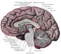
 Pineal gland or epiphysis (in red) | |
 Diagram of pituitary and pineal glands in the human brain | |
| Details | |
| Precursor | Neural ectoderm, roof of diencephalon |
| Artery | Posterior cerebral artery |
| Identifiers | |
| Latin | glandula pinealis |
| MeSH | D010870 |
| NeuroNames | 297 |
| NeuroLex ID | birnlex_1184 |
| TA98 | A11.2.00.001 |
| TA2 | 3862 |
| FMA | 62033 |
| Anatomical terms of neuroanatomy | |
The pineal gland (also known as the pineal body
The pineal gland is present in almost all vertebrates, but is absent in
Ancient Greeks were the first to notice the pineal gland and believed it to be a valve, a guardian for the flow of pneuma. Galen in the 2nd century C.E. could not find any functional role and regarded the gland as a structural support for the brain tissue. He gave the name konario, meaning cone or pinecone, which during Renaissance was translated to Latin as pinealis. In the 17th century, René Descartes revived the mystical purpose and described the gland as the "principal seat of the soul". In the mid-20th century, the real biological role as a neuroendocrine organ was established.[9]
Etymology
The word pineal, from Latin pinea (pine-cone) in reference to the gland's similar shape, was first used in the late 17th century.[3]
Structure
The pineal gland is a pine cone-shaped (hence the name), unpaired midline brain structure.[3][10] It is reddish-gray in colour and about the size of a grain of rice (5–8 mm) in humans. It forms part of the epithalamus.[1] It is attached to the rest of the brain by a pineal stalk.[11] The ventral lamina of the pineal stalk is continuous with the posterior commissure, and its dorsal lamina with the habenular commissure.[11]
Location
It normally lies in a depression between the two
Blood supply
Unlike most of the mammalian brain, the pineal gland is not isolated from the body by the blood–brain barrier system;[13] it has profuse blood flow, second only to the kidney,[14] supplied from the choroidal branches of the posterior cerebral artery.
Afferents
The afferent nerve suppy of pineal gland is by the nervus conarii which receives postganglionic
Neural pathway for melatonin production
The canonical neural pathway regulating pineal melatonin production begins in the eye with the intrinsically photosensitive ganglion cells of the retina which project inhibitory GABAergic efferents to the paraventricular nucleus of hypothalamus via the retinohypothalamic tract. The paraventricular nucleus in turn projects to the superior cervical ganglia, which finally projects to the pineal gland. Darkness thus leads to disinhibition of the paraventricular nucleus, leading it to activate pineal gland melatonin production by way of the superior cervical ganglia.[18]
Microanatomy


The pineal body in humans consists of a lobular
The pineal gland consists mainly of
| Cell type | Description |
|---|---|
| Pinealocytes | The pinealocytes consist of a cell body with 4–6 processes emerging. They produce and secrete melatonin. The pinealocytes can be stained by special silver impregnation methods. Their cytoplasm is lightly basophilic. With special stains, pinealocytes exhibit lengthy, branched cytoplasmic processes that extend to the connective septa and its blood vessels. |
Interstitial cells |
Interstitial cells are located between the pinealocytes. They have elongated nuclei and a cytoplasm that is stained darker than that of the pinealocytes. |
| Perivascular phagocyte | Many capillaries are present in the gland, and perivascular phagocytes are located close to these blood vessels. The perivascular phagocytes are antigen presenting cells. |
| Pineal neurons | In higher vertebrates neurons are usually located in the pineal gland. However, this is not the case in rodents.
|
| Peptidergic neuron-like cells | In some species, neuronal-like peptidergic cells are present. These cells might have a paracrine regulatory function. |
Development
The human pineal gland grows in size until about 1–2 years of age, remaining stable thereafter,[20][21] although its weight increases gradually from puberty onwards.[22][23] The abundant melatonin levels in children are believed to inhibit sexual development, and pineal tumors have been linked with precocious puberty. When puberty arrives, melatonin production is reduced.[24]
Symmetry
In the zebrafish the pineal gland does not straddle the midline, but shows a left-sided bias. In humans, functional cerebral dominance is accompanied by subtle anatomical asymmetry.[25][26][27]
Function
One function of the pineal gland is to produce
The compound
Regulation of the pituitary gland
Studies on rodents suggest that the pineal gland influences the pituitary gland's secretion of the sex hormones, follicle-stimulating hormone (FSH), and luteinizing hormone (LH). Pinealectomy performed on rodents produced no change in pituitary weight, but caused an increase in the concentration of FSH and LH within the gland.[31] Administration of melatonin did not return the concentrations of FSH to normal levels, suggesting that the pineal gland influences pituitary gland secretion of FSH and LH through an undescribed transmitting molecule.[31]
The pineal gland contains
Regulation of bone metabolism
Studies in mice suggest that the pineal-derived melatonin regulates new bone deposition. Pineal-derived melatonin mediates its action on the bone cells through MT2 receptors. This pathway could be a potential new target for osteoporosis treatment as the study shows the curative effect of oral melatonin treatment in a postmenopausal osteoporosis mouse model.[34]
Clinical significance
Calcification
Calcification of the pineal gland is typical in young adults, and has been observed in children as young as two years of age.[35] The internal secretions of the pineal gland are known to inhibit the development of the reproductive glands because when it is severely damaged in children, development of the sexual organs and the skeleton are accelerated.[36] Pineal gland calcification is detrimental to its ability to synthesize melatonin[37][38] and scientific literature presents inconclusive findings on whether it causes sleep problems.[39][40]
The calcified gland is often seen in skull X-rays.[35] Calcification rates vary widely by country and correlate with an increase in age, with calcification occurring in an estimated 40% of Americans by age seventeen.[35] Calcification of the pineal gland is associated with corpora arenacea, also known as "brain sand".
Tumors
A pineal tumor can compress the superior colliculi and pretectal area of the dorsal midbrain, producing Parinaud's syndrome. Pineal tumors also can cause compression of the cerebral aqueduct, resulting in a noncommunicating hydrocephalus. Other manifestations are the consequence of their pressure effects and consist of visual disturbances, headache, mental deterioration, and sometimes dementia-like behaviour.[42]
These
Other conditions
The morphology of the pineal gland differs markedly in different pathological conditions. For instance, it is known that its volume is reduced both in obese patients as well as patients with primary insomnia.[45]
Other animals
Nearly all vertebrate species possess a pineal gland. The most important exception is a primitive vertebrate, the
The results of various scientific research in evolutionary biology, comparative neuroanatomy and neurophysiology have explained the evolutionary history (
During embryonic development, the parietal eye and the pineal organ of modern lizards[55] and tuataras[56] form together from a pocket formed in the brain ectoderm. The loss of parietal eyes in many living tetrapods is supported by developmental formation of a paired structure that subsequently fuses into a single pineal gland in developing embryos of turtles, snakes, birds, and mammals.[57]
The pineal organs of mammals fall into one of three categories based on shape. Rodents have more structurally complex pineal glands than other mammals.[58]
All amphibians have a pineal organ, but some frogs and toads also have what is called a "frontal organ", which is essentially a parietal eye.[61]
Pinealocytes in many non-mammalian vertebrates have a strong resemblance to the photoreceptor cells of the eye. Evidence from morphology and developmental biology suggests that pineal cells possess a common evolutionary ancestor with retinal cells.[62]
Pineal
In most vertebrates, exposure to light sets off a chain reaction of enzymatic events within the pineal gland that regulates
The fossilized skulls of many extinct vertebrates have a pineal
History

The secretory activity of the pineal gland is only partially understood. Its location deep in the brain suggested to philosophers throughout history that it possesses particular importance. This combination led to its being regarded as a "mystery" gland with
Galen described the pineal gland in De usu partium corporis humani, libri VII (On the Usefulness of Parts of the Body, Part 8) and De anatomicis administrationibus, libri IX (On Anatomical Procedures, Part 9).[71] He introduced the name κωνάριο (konario, often Latinised as conarium) that means cone, as in pinecone,[72] in De usu partium corporis humani. He correctly located the gland as directly lying behind the third ventricle. He argued against the prevailing concept as a valve for two basic reasons: it is located outside of the brain tissue and it does not move on its own.[67]
Galen instead identified the valve as a worm-like structure in the
Seventeenth-century philosopher and scientist René Descartes discussed the pineal gland both in his first book, the Treatise of Man (written before 1637, but only published posthumously 1662/1664), and in his last book, The Passions of the Soul (1649) and he regarded it as "the principal seat of the soul and the place in which all our thoughts are formed".[9] In the Treatise of Man, he described conceptual models of man, namely creatures created by God, which consist of two ingredients, a body and a soul.[9][76] In the Passions, he split man up into a body and a soul and emphasized that the soul is joined to the whole body by "a certain very small gland situated in the middle of the brain's substance and suspended above the passage through which the spirits in the brain's anterior cavities communicate with those in its posterior cavities". Descartes gave importance to the structure as it was the only unpaired component of the brain.[9]
The Latin name pinealis became popular in the 17th century. For example, English physician Thomas Willis described glandula pinealis book, Cerebri anatome cui accessit nervorum descriptio et usus (1664). Willis criticised Descartes' concept, remarking: "we can scarce[ly] believe this to be the seat of the Soul, or its chief Faculties to arise from it; because Animals, which seem to be almost quite destitute of Imagination, Memory, and other superior Powers of the Soul, have this Glandula or Kernel large and fair enough."[75]
The pineal gland was originally believed to be a "
Society and culture
The notion of a "pineal-eye" is central to the philosophy of the French writer Georges Bataille, which is analyzed at length by literary scholar Denis Hollier in his study Against Architecture. In this work Hollier discusses how Bataille uses the concept of a "pineal-eye" as a reference to a blind-spot in Western rationality, and an organ of excess and delirium.[84] This conceptual device is explicit in his surrealist texts, The Jesuve and The Pineal Eye.[85]
In the late 19th century
In the short story "From Beyond" by H. P. Lovecraft, a scientist creates an electronic device that emits a resonance wave, which stimulates an affected person's pineal gland, thereby allowing them to perceive planes of existence outside the scope of accepted reality, a translucent, alien environment that overlaps our own recognized reality. It was adapted as a film of the same name in 1986. The 2013 horror film Banshee Chapter is heavily influenced by this short story. In Season 16, episode 6 of "American Dad" Steve tries to "astral project" using his pineal gland to help him understand the meaning of life. The episode is entitled "The Wondercabinet".
Additional images
The pineal body is labeled in these images.
-
 Mesal aspect of a brain sectioned in the median sagittal plane
 -
 Dissection showing the ventricles of the brain
 -
 Hind- and mid-brains; antero-lateral view
 -
 Median sagittal section of brain
 -
 Pineal gland
 -
 Brainstem; posterior view


See also
References
- ^ PMID 9802483.
- PMID 1778647.
- ^ a b c "Pineal (as an adjective)". Online Etymology Dictionary, Douglas Harper. 2018. Retrieved 27 October 2018.
- S2CID 26142713.
- PMID 15649736.
Exogenous melatonin has acute sleepiness-inducing and temperature-lowering effects during 'biological daytime', and when suitably timed (it is most effective around dusk and dawn) it will shift the phase of the human circadian clock (sleep, endogenous melatonin, cortisol) to earlier (advance phase shift) or later (delay phase shift) times.
- S2CID 18748366.
- ^ S2CID 40006945.
- ^ a b Eakin RM (1973). The Third Eye. Berkeley: University of California Press.
- ^ a b c d e Lokhorst GJ (2018). Zalta EN (ed.). "Descartes and the Pineal Gland; In: The Stanford Encyclopedia of Philosophy" (Winter 2018 ed.). Metaphysics Research Lab, Stanford University. Retrieved 17 December 2019.
- ^ Bowen R. "The Pineal Gland and Melatonin". Archived from the original on 24 November 2011. Retrieved 14 October 2011.
- ^ ISBN 978-0-07-160399-7.
- ISBN 978-1-4160-6257-8.
- ISBN 978-1-889325-29-3. Retrieved 8 February 2009.
- ^ Arendt J: Melatonin and the Mammalian Pineal Gland, ed 1. London. Chapman & Hall, 1995, p 17
- ^ S2CID 25719864.
- ^ S2CID 40328108.
- S2CID 5763932.
- PMID 33897495.
- PMID 17076524.
- PMID 7536203.
- PMID 8938291.
- S2CID 38346296.
- S2CID 28529644.
- ^ "Melatonin Production and Age". Chronobiology. Medichron Publications.
- PMID 18385257.
- PMID 18629869.
- PMID 19084075.
- PMID 4195878.
- PMID 11092838.
- .
- ^ S2CID 28964258.
- S2CID 39053816.
- S2CID 12713010.
- PMID 28512916.
- ^ PMID 7063680. Archived from the original(PDF) on 24 March 2012. Retrieved 21 June 2012.
- ^ "The Pineal Body". Human Anatomy (Gray's Anatomy). Archived from the original on 26 August 2011. Retrieved 7 September 2011.
- S2CID 20184373.
- PMID 29385085.
- PMID 25277665.
- PMID 18755628.
- ISBN 9780323296359.
- ^ Bruce J. "Pineal Tumours". eMedicine. Archived from the original on 5 September 2015. Retrieved 25 September 2015.
- ^ "Pineal Tumours". American Brain Tumour Association. Archived from the original on 26 September 2015. Retrieved 25 September 2015.
- PMID 20461445.
- S2CID 25611663.
- S2CID 29929205.
- ^ PMID 9514841.
- S2CID 25837046.
- PMID 23575111.
- PMID 7148728.
- ISBN 978-3-642-65497-8, retrieved 28 March 2023
- S2CID 85280810.
- ISBN 9780226315683. Retrieved 2 February 2017.
- ^ Dodt E (1973). "The parietal eye (pineal and parietal organs) of lower vertebrates". Visual Centers in the Brain. Springer. pp. 113–140.
- .
- .
- ^ a b Quay WB (1979). "The parietal eye–pineal complex". In Gans C, Northcutt RG, Ulinski P (eds.). Biology of the Reptilia. Volume 9. Neurology A. London: Academic Press. pp. 245–406. Archived from the original on 3 February 2017.
- ^ PMID 398532.
- ^ S2CID 30406445.
- .
- S2CID 33692776.
- S2CID 26023207.
- S2CID 44377291.
- ^ Edinger T (1955). "The size of parietal foramen and organ in reptiles: a rectification". Bulletin of the Museum of Comparative Zoology. 114: 1–34. Archived from the original on 1 December 2017.
- PMID 17426009.
- ^ S2CID 26207380.
- PMID 21801980.
- S2CID 9140909.
- ISBN 978-3-319-41678-6.
- ISBN 978-81-322-2801-1. Retrieved 28 March 2023.
- ^ PMID 29278521.
- S2CID 255470624.
- PMID 15300863.
- ^ ISSN 2155-3017. Retrieved 28 March 2023.
- ISBN 978-0-19-514581-6.
- ^ Spencer B (1885). "On the Presence and Structure of the Pineal Eye in Lacertilia". Quarterly Journal of Microscopy. London: Bartholomew Close. pp. 1–76.
- ^ Flemming AF (1991). "A third eye". Culna (40): 26–27 – via Sabinet.
- ISBN 978-0-520-32632-3.
- ^ PMID 14298722.
- PMID 14415935.
- ISBN 978-0-8247-5504-1. Retrieved 31 March 2009.
- .
- ^ Hollier D (1989). Against Architecture: The Writings of Georges Bataille. Translated by Wing B. MIT.
- ^ Bataille G (1985). Visions of excess: Selected writings 1927-1939. Manchester University Press.
- ^ "Principia Discordia - Page 15". principiadiscordia.com. Retrieved 22 September 2023.