
Pituitary gland
This article may be too technical for most readers to understand. (September 2023) |
| Pituitary gland | |
|---|---|
inferior hypophyseal artery, capsular artery, artery of the inferior cavernous sinus[1] | |
| Identifiers | |
| Latin | hypophysis, glandula pituitaria |
| MeSH | D010902 |
| NeuroLex ID | birnlex_1353 |
| TA98 | A11.1.00.001 |
| TA2 | 3853 |
| FMA | 13889 |
| Anatomical terms of neuroanatomy] | |


The pituitary gland (or hypophysis cerebri) is an endocrine gland in vertebrates. In humans, the pituitary gland is located at the base of the brain, protruding off the bottom of the hypothalamus. The human pituitary gland is oval shaped, about the size of a chickpea,[2] and weighs 0.5 grams (0.018 oz) on average.
Structure
In humans, the pituitary gland rests upon the
The pituitary gland is composed of the

The height of the pituitary gland ranges from 5.3 to 7.0 mm. The volume of the pituitary gland ranges from 200 to 440 mm3.[6]
Anterior
The anterior pituitary lobe (or adenohypophysis) arises from an invagination of the oral ectoderm (Rathke's pouch). This contrasts with the posterior pituitary, which originates from neuroectoderm.
The anterior lobe of the pituitary can be divided into the
The anterior pituitary contains several different types of cells[8] that synthesize and secrete hormones. Usually there is one type of cell for each major hormone formed in anterior pituitary. With special stains attached to high-affinity antibodies that bind with distinctive hormone, at least 5 types of cells can be differentiated.
| S.No. | Type of cell | Hormone secreted | Percentage of type of cell |
|---|---|---|---|
| 1. | Somatotropes | human Growth Hormone (hGH) | 30–50% |
| 2. | Corticotropes | AdrenoCorticoTropic Hormone (ACTH) | 20% |
| 3. | Thyrotropes | Thyroid-Stimulating Hormone (TSH) | 3–5% |
| 4. | Gonadotropes | Gonadotropic hormones = both Luteinizing Hormone (LH) and Follicle-Stimulating Hormone (FSH) | 3–5% |
| 5. | Lactotropes | Prolactin (PRL) | 3–5% |
Posterior
The posterior lobe develops as an extension of the hypothalamus, from the floor of the third ventricle. The posterior pituitary hormones are synthesized by cell bodies in the hypothalamus. The magnocellular neurosecretory cells, of the supraoptic and paraventricular nuclei located in the hypothalamus, project axons down the infundibulum to terminals in the posterior pituitary. This simple arrangement differs sharply from that of the adjacent anterior pituitary, which does not develop from the hypothalamus.
The release of pituitary hormones by both the anterior and posterior lobes is under the control of the hypothalamus, albeit in different ways.[7]
Functions
Anterior
The anterior pituitary synthesizes and secretes hormones. All releasing hormones (-RH) referred to can also be referred to as releasing factors (-RF).
- growth hormone-releasing hormone (GHRH), and is inhibited by hypothalamic somatostatin.
- Cleaved from the precursor beta-endorphin, and melanocyte-stimulating hormone are released under the influence of hypothalamic corticotropin-releasing hormone (CRH).[9][10]: 1210
- Thyroid-stimulating hormone (TSH) is released under the influence of hypothalamic thyrotropin-releasing hormone (TRH) and is inhibited by somatostatin.
- Luteinizing hormone (LH).
- Follicle-stimulating hormone (FSH), both released under influence of Gonadotropin-releasing Hormone (GnRH)
- Prolactin (PRL), whose release is inconsistently stimulated by hypothalamic TRH, oxytocin, vasopressin, vasoactive intestinal peptide, angiotensin II, neuropeptide Y, galanin, substance P, bombesin-like peptides (gastrin-releasing peptide, neuromedin B and C), and neurotensin, and inhibited by hypothalamic dopamine.[11]
These hormones are released from the anterior pituitary under the influence of the hypothalamus. Hypothalamic hormones are secreted to the anterior lobe by way of a special capillary system, called the hypothalamic-hypophysial portal system.
There is also a non-endocrine cell population called folliculostellate cells.
Intermediate
The intermediate lobe synthesizes and secretes the following important endocrine hormone:
- Melanocyte–stimulating hormone (MSH). This is also produced in the anterior lobe.[12] When produced in the intermediate lobe, MSHs are sometimes called "intermedins".
Posterior
The posterior pituitary stores and secretes (but does not synthesize) the following important endocrine hormones:
- Antidiuretic hormone (ADH, also known as vasopressin and arginine vasopressin AVP), the majority of which is released from the supraoptic nucleus in the hypothalamus.
- paraventricular nucleus in the hypothalamus. Oxytocin is one of the few hormones to create a positive feedbackloop. For example, uterine contractions stimulate the release of oxytocin from the posterior pituitary, which, in turn, increases uterine contractions. This positive feedback loop continues throughout labour.
Hormones
Hormones secreted from the pituitary gland help control the following body processes:
- Growth(GH)
- Blood pressure
- Some aspects of pregnancy and childbirth including stimulation of uterine contractions
- Breast milk production
- Sex organ functions in both sexes
- Thyroid glandfunction
- Metabolic conversion of food into energy
- osmolarityregulation in the body
- Water balance via the control of reabsorption of kidneys
- Temperature regulation
- Pain relief
Clinical significance

Some of the diseases involving the pituitary gland are:
- Central diabetes insipidus caused by a deficiency of vasopressin
- Gigantism and acromegaly caused by an excess of growth hormone in childhood and adult, respectively
- Hypothyroidism caused by a deficiency of thyroid-stimulating hormone
- Hyperpituitarism, the increased (hyper) secretion of one or more of the hormones normally produced by the pituitary gland
- Hypopituitarism, the decreased (hypo) secretion of one or more of the hormones normally produced by the pituitary gland
- Panhypopituitarisma decreased secretion of most of the pituitary hormones
- Pituitary tumours
- tumorsthat occur in the pituitary gland
All of the functions of the pituitary gland can be adversely affected by an over- or under-production of associated hormones.
The pituitary gland is important for mediating the stress response, via the hypothalamic–pituitary–adrenal axis (HPA axis). Critically, pituitary gland growth during adolescence can be altered by early life stress such as childhood maltreatment or maternal dysphoric behavior.[13]
It has been demonstrated that, after controlling for age, sex, and BMI, larger quantities of
History
Etymology
Pituitary gland
The Greek
The expression glandula pituitaria is still used as official synonym beside hypophysis in the official Latin nomenclature Terminologia Anatomica.[20] In the seventeenth century the supposed function of the pituitary gland to produce nasal mucus was debunked.[15] The expression glandula pituitaria and its English equivalent pituitary gland can only be justified from a historical point of view.[21] The inclusion of this synonym is merely justified by noting that the main term hypophysis is a much less popular term.[22]
Hypophysis
Note: hypophysial (or hypophyseal) means "related to the hypophysis (pituitary gland)".
The anatomist Samuel Thomas von Sömmerring coined the name hypophysis.[15] This name consists[15][21] of ὑπό ('under')[16] and φύειν ('to grow').[16] In later Greek ὑπόφυσις is used differently by Greek physicians as outgrowth.[15] Sömmering also used the equivalent expression appendix cerebri,[15][18] with appendix as appendage.[17] In various languages, Hirnanhang[18] in German and hersenaanhangsel[23] in Dutch, the terms are derived from appendix cerebri.
Other animals
The pituitary gland is found in all vertebrates, but its structure varies among different groups.
The division of the pituitary described above is typical of mammals, and is also true, to varying degrees, of all tetrapods. However, only in mammals does the posterior pituitary have a compact shape. In lungfish, it is a relatively flat sheet of tissue lying above the anterior pituitary, but in amphibians, reptiles, and birds, it becomes increasingly well developed. The intermediate lobe is, in general, not well developed in any species and is entirely absent in birds.[24]
The structure of the pituitary in fish, apart from the lungfish, is generally different from that in other animals. In general, the intermediate lobe tends to be well developed, and may equal the remainder of the anterior pituitary in size. The posterior lobe typically forms a sheet of tissue at the base of the pituitary stalk, and in most cases sends irregular finger-like projection into the tissue of the anterior pituitary, which lies directly beneath it. The anterior pituitary is typically divided into two regions, a more anterior rostral portion and a posterior proximal portion, but the boundary between the two is often not clearly marked. In
The arrangement in lampreys, which are among the most primitive of all fish, may indicate how the pituitary originally evolved in ancestral vertebrates. Here, the posterior pituitary is a simple flat sheet of tissue at the base of the brain, and there is no pituitary stalk. Rathke's pouch remains open to the outside, close to the nasal openings. Closely associated with the pouch are three distinct clusters of glandular tissue, corresponding to the intermediate lobe, and the rostral and proximal portions of the anterior pituitary. These various parts are separated by meningial membranes, suggesting that the pituitary of other vertebrates may have formed from the fusion of a pair of separate, but associated, glands.[24]
Most
There is a structure analogous to the pituitary in the octopus brain.[25]
Intermediate lobe
Although rudimentary in humans (and often considered part of the anterior pituitary), the intermediate lobe located between the anterior and posterior pituitary is important to many animals. For instance, in fish, it is believed to control physiological color change. In adult humans, it is just a thin layer of cells between the anterior and posterior pituitary. The intermediate lobe produces melanocyte-stimulating hormone (MSH), although this function is often (imprecisely) attributed to the anterior pituitary.
The intermediate lobe is, in general, not well developed in tetrapods, and is entirely absent in birds.[24]
-
 Location of the pituitary gland in the human brain
 -
 Pituitary and pineal glands
 -
 The arteries of the base of the brain
 -
 Mesal aspect of a brain sectioned in the median sagittal plane
 -
 Pituitary
 -
 Pituitary gland
 -
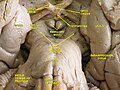 Cerebrum. Inferior view. Deep dissection.


See also
References
- PMID 8254920.
- ^ Leng, Gareth (2018). The Heart of the Brain: The Hypothalamus and its Hormones.
The gland in humans is described in Wikipedia as being the size of a pea. So common is this description that it seemed likely to be wrong, as I confirmed by examining a selection of peas. Wikipedia also gives the weight of the human pituitary as about half a gram, and in this it is more correct. The pituitary in a human is at least five times the average size of my peas; it is more like the size of a chickpea.
- ISBN 978-1-4377-3580-2.
- PMID 31855373. Retrieved 24 Sep 2020.
- ^ ISBN 978-0-12-380926-1.
- .
- ^ ISBN 978-1-4160-3115-4.
- ^ Textbook of Medical Physiology. Elsevier Saunders.
- PMID 6328345.
- ISBN 978-0-07-162442-8.
- ISBN 978-0-12-380926-1.
- ISBN 978-0-19-856878-0.
- S2CID 5247274.
- ^ S2CID 22520320.
- ^ a b c d e f g h Hyrtl, J. (1880). Onomatologia Anatomica. Geschichte und Kritik der anatomischen Sprache der Gegenwart. Wien: Wilhelm Braumüller. K.K. Hof- und Universitätsbuchhändler.
- ^ a b c Liddell, H.G. & Scott, R. (1940). A Greek-English Lexicon. revised and augmented throughout by Sir Henry Stuart Jones. with the assistance of. Roderick McKenzie. Oxford: Clarendon Press.
- ^ a b Lewis, C.T. & Short, C. (1879). A Latin dictionary founded on Andrews' edition of Freund's Latin dictionary. Oxford: Clarendon Press.
- ^ a b c Schreger, C.H.Th.(1805). Synonymia anatomica. Synonymik der anatomischen Nomenclatur. Fürth: im Bureau für Literatur.
- ^ Anderson, D.M. (2000). Dorland's illustrated medical dictionary (29th edition). Philadelphia/London/Toronto/Montreal/Sydney/Tokyo: W.B. Saunders Company.
- ^ Federative Committee on Anatomical Terminology (FCAT) (1998). Terminologia Anatomica. Stuttgart: Thieme
- ^ a b Triepel, H. (1927). Die anatomischen Namen. Ihre Ableitung und Aussprache. Anhang: Biographische Notizen.(Elfte Auflage). München: Verlag J.F. Bergmann.
- ^ International Anatomical Nomenclature Committee (1966). Nomina Anatomica. Amsterdam: Excerpta Medica Foundation, p. 62
- ^ Pinkhof, H. (1923). Vertalend en verklarend woordenboek van uitheemsche geneeskundige termen. Haarlem: De Erven F. Bohn.
- ^ ISBN 0-03-910284-X.
- S2CID 4159935.
External links
- hier-382 at NeuroNames
- Histology image: 14201loa – Histology Learning System at Boston University
- The Pituitary Gland, from the UMM Endocrinology Health Guide Archived 2010-06-20 at the Wayback Machine
- Oklahoma State, Endocrine System
- The Pituitary Foundation
- The Pituitary Network Association -- pituitary.org