
Placenta
| Placenta | |
|---|---|
chorion frondosum | |
| Identifiers | |
| Latin | placenta |
| MeSH | D010920 |
| TE | E5.11.3.1.1.0.5 |
| Anatomical terminology] | |
The placenta (pl.: placentas or placentae) is a temporary
The word placenta comes from the Latin word for a type of cake, from Greek πλακόεντα/πλακοῦντα plakóenta/plakoúnta, accusative of πλακόεις/πλακούς plakóeis/plakoús, "flat, slab-like",[6][7] with reference to its round, flat appearance in humans. The classical plural is placentae, but the form placentas is more common in modern English.
Evolution and phylogenetic diversity
The placenta has evolved independently multiple times, probably starting in fish, where it originated multiple times, including the genus Poeciliopsis.[8] Placentation has also evolved in some reptiles.[9]
The mammalian placenta evolved more than 100 million years ago and was a critical factor in the explosive diversification of placental mammals.[10] Although all mammalian placentas have the same functions, there are important differences in structure and function in different groups of mammals. For example, human, bovine, equine and canine placentas are very different at both the gross and the microscopic levels. Placentas of these species also differ in their ability to provide maternal immunoglobulins to the fetus.[11]
Structure
Placental mammals, including humans, have a chorioallantoic placenta that forms from the
The placenta occasionally takes a form in which it comprises several distinct parts connected by blood vessels.[14] The parts, called lobes, may number two, three, four, or more. Such placentas are described as bilobed/bilobular/bipartite, trilobed/trilobular/tripartite, and so on. If there is a clearly discernible main lobe and auxiliary lobe, the latter is called a succenturiate placenta. Sometimes the blood vessels connecting the lobes get in the way of fetal presentation during labor, which is called vasa previa.[citation needed]
Gene and protein expression
About 20,000 protein coding genes are expressed in human cells and 70% of these genes are expressed in the normal mature placenta.
Physiology
Development

The placenta begins to develop upon implantation of the blastocyst into the maternal endometrium, very early on in pregnancy at about week 4.[17]
The outer layer of the late blastocyst, is formed of trophoblasts, cells that form the outer layer of the placenta. This outer layer is divided into two further layers: the underlying cytotrophoblast layer and the overlying syncytiotrophoblast layer. The syncytiotrophoblast is a multinucleated continuous cell layer that covers the surface of the placenta. It forms as a result of differentiation and fusion of the underlying cytotrophoblasts, a process that continues throughout placental development. The syncytiotrophoblast contributes to the barrier function of the placenta.[18]
The placenta grows throughout pregnancy. Development of the maternal blood supply to the placenta is complete by the end of the first trimester of pregnancy week 14 (DM).[17]
Placental circulation

Maternal placental circulation
In preparation for implantation of the blastocyst, the endometrium undergoes
Maternal blood flow begins between days 5–12,[19] and is approximately 600–700 ml/min at term.
Fetoplacental circulation
Deoxygenated fetal blood passes through
Endothelin and prostanoids cause vasoconstriction in placental arteries, while nitric oxide causes vasodilation.[21] On the other hand, there is no neural vascular regulation, and catecholamines have only little effect.[21]
The fetoplacental circulation is vulnerable to persistent hypoxia or intermittent hypoxia and reoxygenation, which can lead to generation of excessive
This begins at day 17–22.[23]
Birth
Placental expulsion begins as a physiological separation from the wall of the uterus. The period from just after the child is born until just after the placenta is expelled is called the "third stage of labor". The placenta is usually expelled within 15–30 minutes of birth.[citation needed]
Placental expulsion can be managed actively, for example by giving oxytocin via intramuscular injection followed by cord traction to assist in delivering the placenta. Alternatively, it can be managed expectantly, allowing the placenta to be expelled without medical assistance. Blood loss and the risk of postpartum bleeding may be reduced in women offered active management of the third stage of labour, however there may be adverse effects and more research is necessary.[24]
The habit is to cut the cord immediately after birth, but it is theorised that there is no medical reason to do this; on the contrary, it is theorised that not cutting the cord helps the baby in its adaptation to extrauterine life, especially in preterm infants.[25]
Microbiome
The placenta is traditionally thought to be
Functions
Nutrition and gas exchange
The placenta intermediates the transfer of nutrients between mother and fetus. The perfusion of the intervillous spaces of the placenta with maternal blood allows the transfer of nutrients and oxygen from the mother to the fetus and the transfer of waste products and carbon dioxide back from the fetus to the maternal blood. Nutrient transfer to the fetus can occur via both active and passive transport.[30] Placental nutrient metabolism was found to play a key role in limiting the transfer of some nutrients.[31] Adverse pregnancy situations, such as those involving maternal diabetes or obesity, can increase or decrease levels of nutrient transporters in the placenta potentially resulting in overgrowth or restricted growth of the fetus.[32]
Excretion
Waste products excreted from the fetus such as urea, uric acid, and creatinine are transferred to the maternal blood by diffusion across the placenta.[citation needed]
Immunity
The placenta functions as a selective barrier between maternal and fetal cells, preventing maternal blood, proteins and
Beginning as early as 13 weeks of gestation, and increasing linearly, with the largest transfer occurring in the third trimester,
Endocrine function
- The first hormone released by the placenta is called the human chorionic gonadotropin (hCG) hormone. This is responsible for stopping the process at the end of menses when the corpus luteum ceases activity and atrophies. If hCG did not interrupt this process, it would lead to spontaneous abortion of the fetus. The corpus luteum also produces and releases progesterone and estrogen, and hCG stimulates it to increase the amount that it releases. hCG is the indicator of pregnancy that pregnancy tests look for. These tests will work when menses has not occurred or after implantation has happened on days seven to ten. hCG may also have an anti-antibody effect, protecting it from being rejected by the mother's body. hCG also assists the male fetus by stimulating the testes to produce testosterone, which is the hormone needed to allow the sex organs of the male to grow.
- Progesterone helps the embryo implant by assisting passage through the fallopian tubes. It also affects the fallopian tubes and the uterus by stimulating an increase in secretions necessary for fetal nutrition. Progesterone, like hCG, is necessary to prevent spontaneous abortion because it prevents contractions of the uterus and is necessary for implantation.
- Estrogen is a crucial hormone in the process of proliferation. This involves the enlargement of the breasts and uterus, allowing for growth of the fetus and production of milk. Estrogen is also responsible for increased blood supply towards the end of pregnancy through vasodilation. The levels of estrogen during pregnancy can increase so that they are thirty times what a non-pregnant woman mid-cycles estrogen level would be.
Immunological barrier
The placenta and fetus may be regarded as a foreign body inside the mother and must be protected from the normal immune response of the mother that would cause it to be rejected. The placenta and fetus are thus treated as sites of immune privilege, with immune tolerance.
For this purpose, the placenta uses several mechanisms :
- It secretes
- There is presence of small lymphocytic suppressor cells in the fetus that inhibit maternal cytotoxic T cells by inhibiting the response to interleukin 2.[45]
However, the placental barrier is not the sole means of evading the immune system, as foreign fetal cells also persist in the maternal circulation, on the other side of the placental barrier.[46]
DNA methylation
The trophoblast is the outer layer of cells of the blastocyst (see day 9 in Figure, above, showing the initial stages of human embryogenesis). Placental trophoblast cells have a unique genome-wide DNA methylation pattern determined by de novo methyltransferases during embryogenesis.[47] This methylation pattern is principally required to regulate placental development and function, which in turn is critical for embryo survival.[47]
Other
The placenta also provides a reservoir of blood for the fetus, delivering blood to it in case of hypotension and vice versa, comparable to a capacitor.[48]

Clinical significance

Numerous pathologies can affect the placenta.[citation needed]
- Placenta accreta, when the placenta implants too deeply, all the way to the actual muscle of uterine wall (without penetrating it)
- Placenta praevia, when the placement of the placenta is too close to or blocks the cervix
- Placental abruption, premature detachment of the placenta
- TORCH infections.
Society and culture
The placenta often plays an important role in various
Some cultures
The placenta is believed by some communities to have power over the lives of the baby or its parents. The
Several cultures believe the placenta to be or have been alive, often a relative of the baby. Nepalese think of the placenta as a friend of the baby; the orang Asli and Malay populations in Malay Peninsula regard it as the baby's older sibling.[53][55] Native Hawaiians believe that the placenta is a part of the baby, and traditionally plant it with a tree that can then grow alongside the child.[49] Various cultures in Indonesia, such as Javanese and Malay, believe that the placenta has a spirit and needs to be buried outside the family house. Some Malays would bury the baby's placenta with a pencil (if it is a boy) or a needle and thread (if it is a girl).[55]
In some cultures, the placenta is eaten, a practice known as placentophagy. In some eastern cultures, such as China, the dried placenta (ziheche 紫河车, literally "purple river car") is thought to be a healthful restorative and is sometimes used in preparations of traditional Chinese medicine and various health products.[56] The practice of human placentophagy has become a more recent trend in western cultures and is not without controversy; its practice being considered cannibalism is debated.
Some cultures have alternative uses for placenta that include the manufacturing of cosmetics, pharmaceuticals and food.[57]
Additional images
-
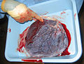 Human placenta immediately post birth.
 -
 Fetus of about 8 weeks, enclosed in the amnion. Magnified a little over two diameters.
 -
 Placenta with attached fetal membranes, ruptured at the margin at the left in the image
 -
-
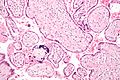
 Micrograph of CMV placentitis
 -
 A 3D Power Doppler image of vasculature in 20-week placenta
 -
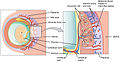 Schematic view of the placenta
 -
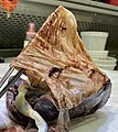
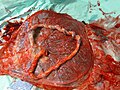
 Maternal side of a whole human placenta, just after birth
 -
 Fetal side of same placenta
 -
 Close-up of umbilical attachment to fetal side of freshly delivered placenta
 -
![Placenta weight by gestational age[58]](//upload.wikimedia.org/wikipedia/commons/thumb/5/5b/Placenta_weight_by_gestational_age.svg/120px-Placenta_weight_by_gestational_age.svg.png) Placenta weight by gestational age[58]
Placenta weight by gestational age[58] -
 Ziheche (紫河车), dried human placenta used in traditional Chinese medicine


![Placenta weight by gestational age[58]](/File:Placenta_weight_by_gestational_age.svg)

See also
References
- PMID 36144676.
- ISBN 978-0-13-100849-6.[page needed]
- ^ Mitra A (31 January 2020). "How the placenta evolved from an ancient virus". WHYY. Retrieved 9 March 2020.
- PMID 30300353.
- S2CID 12042851.
- ^ Henry George Liddell, Robert Scott, "A Greek-English Lexicon", at Perseus Archived 2012-04-05 at the Wayback Machine.
- ^ "placenta" Archived 2016-01-30 at the Wayback Machine. Online Etymology Dictionary.
- S2CID 2372572.
- PMID 26474917.
- PMID 17494753.
- ^ Bowen R. "Implantation and Development of the Placenta: Introduction and Index". Pathophysiology of the Reproductive System. Colorado State. Retrieved 7 July 2019.
- PMID 9518951. Archived from the originalon 2011-10-16.
- ^ "Placental Structure and Classification". Colorado State. Archived from the original on 2016-02-11.
- PMID 5429965.
Bipartite placenta represented 4.2 per cent (366 of 8,505) of placentas of white women at the Boston Hospital for Women who were enrolled in the Collaborative Project.
- ^ "The human proteome in placenta - The Human Protein Atlas". www.proteinatlas.org. Archived from the original on 2017-09-26. Retrieved 2017-09-26.
- S2CID 802377.
- ^ a b "Stages of Development of the Fetus - Women's Health Issues". Merck Manuals Consumer Version. Retrieved 2022-06-12.
- ^ "How Your Fetus Grows During Pregnancy". www.acog.org. Retrieved 2022-06-12.
- ISBN 978-1-259-64433-7.[page needed]
- ^ "Placental blood circulation". Online course in embryology for medical students. Universities of Fribourg, Lausanne and Bern (Switzerland). Archived from the original on 2011-09-28.
- ^ S2CID 25040285.
- ^ PMID 24132226.
- ^ Williams book of obsteritcis.
- PMID 30754073.
- PMID 19847185.
- PMID 28454555.
- PMID 26428492.
- PMID 24793619.
- PMID 28120852.
- ISBN 978-1-4443-3366-4.
- PMID 27913585.
- PMID 22701643.
- ^ .
- PMID 23298207.
- S2CID 51724379.
- PMID 28494237.
- PMID 22169833.
- PMID 22235228.
- PMID 9501287.
- ISBN 978-1-58255-999-5.
- ^ "What infections can affect pregnancy?". National Institute of Child Health and Human Development. 27 April 2021. Retrieved 25 June 2021.
- S2CID 28778529.
- ^ "Human Placental Lactogen". www.ucsfhealth.org. May 17, 2009. Archived from the original on April 29, 2017. Retrieved July 21, 2017.
- ^ "Placenta 'fools body's defences'". BBC News. 10 November 2007. Archived from the original on 29 April 2012.
- S2CID 22815679.
- PMID 18384774.
- ^ a b Andrews S, Krueger C, Mellado-Lopez M, Hemberger M, Dean W, Perez-Garcia V, Hanna CW. Mechanisms and function of de novo DNA methylation in placental development reveals an essential role for DNMT3B. Nat Commun. 2023 Jan 23;14(1):371. doi: 10.1038/s41467-023-36019-9. PMID: 36690623; PMCID: PMC9870994
- S2CID 8785947.
- ^ a b "Why eat a placenta?". BBC News. 18 April 2006.
- .
- ^ Francisco E (3 December 2004). "Bridging the Cultural Divide in Medicine". Science.
- PMID 11614175.
- ^ a b Buckley SJ. "Placenta Rituals and Folklore from around the World". Mothering. Archived from the original on 6 January 2008. Retrieved 7 January 2008.
- ^ Davenport A (June 2005). "The Love Offer". Johns Hopkins Magazine. 57 (3).
- ^ ISBN 978-967-13305-9-3.
- ^ Falcao R. "Medicinal Uses of the Placenta". Archived from the original on 5 December 2008. Retrieved 25 November 2008.
- S2CID 151874106.
- ISBN 978-1-316-84861-6.
External links
- The placenta-specific proteome at the Human Protein Atlas
- The Placenta, gynob.com, with quotes from Williams Obstetrics, 18th Edition, F. Gary Cunningham, M.D., Paul C. MacDonald, M.D., Norman F. Grant, M.D., Appleton & Lange, Publishers.
| International | |
|---|---|
| National | |