
Pneumocephalus
| Pneumocephalus | |
|---|---|
 | |
| Pneumocephalus and comminuted fracture of the frontal sinus |
Pneumocephalus is the presence of air or gas within the
, and rarely, spontaneously. Pneumocephalus can occur in scuba diving, but is very rare in this context.If there is a valve mechanism which allows air to enter the skull but prevents it from escaping, a tension pneumocephalus can occur (similar to what can happen in a
tension pneumothorax
).
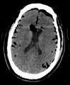
CT scans of patients with a tension pneumocephalus typically show air that compresses the frontal lobes of the brain, which results in a tented appearance of the brain in the skull known as the Mount Fuji sign.[1][2][3] The name is derived from the resemblance of the brain to
bridging veins.[3] Its occurrence seems to be limited to tension pneumocephalus (not occurring in pneumocephalus without tension).[4] The sign was first described by a team of Japanese neurosurgeons.[5]
Pneumocephalus has also been shown to follow neurosurgical procedures such as deep brain stimulation and hematoma evacuation (e.g.,
chronic subdural hematoma[6]), where while seemingly innocuous to the patient, may cause brain shift, subsequent stereotactic inaccuracy, and even another surgical intervention.[7][8] Regarding chronic subdural hematoma (CSDH) surgery, a postoperative volume of pneumocephalus greater than 15mL puts a patient at increased risk of CSDH recurrence; in fact, for every milliliter of air entering the cranial cavity after CSDH evacuation, the recurrence risk increases by 4%.[9]
Efforts are made by neurosurgeons to reduce pneumocephalus volume during surgery, and thus, subsequent brain shift.
Additional images
-
 Large pneumocephaly secondary to surgical wound
 -
 Pneumocephaly

