
Posterior tibial artery
| Posterior tibial artery | |
|---|---|
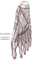
 Back of left lower extremity, showing surface markings for bones, vessels, and nerves (posterior tibial artery labeled at bottom right). | |
| Details | |
| Source | Popliteal artery |
| Branches | Fibular artery, medial plantar artery, lateral plantar artery |
| Vein | Posterior tibial vein |
| Identifiers | |
| Latin | arteria tibialis posterior |
| TA98 | A12.2.16.055 |
| TA2 | 4721 |
| FMA | 43895 |
| Anatomical terminology | |
The posterior tibial artery of the
.Structure
The posterior tibial artery arises from the
medial malleolus of the tibia, but anterior to the Achilles tendon.[1] It passes into the foot deep to the flexor retinaculum of the foot.[1] It runs through the tarsal tunnel.[2]
Branches
The posterior tibial artery gives rise to:
- medial plantar artery.
- lateral plantar artery.
- fibular artery,[1] which is said to rise from the bifurcation of the tibial-fibular trunk and the posterior tibial artery.
- calcaneal branch to the medial aspect of the calcaneus.
Function
The posterior tibial artery supplies oxygenated blood to the posterior compartment of the leg and the plantar surface of the foot.[1]
Clinical significance
Palpation of the posterior tibial artery pulse
The posterior tibial artery
peripheral vascular disease. It is very rarely absent in young and healthy individuals.[3] In a study of 547 healthy individuals, only one person did not have a palpable posterior tibial artery.[3] It is easily palpated over Pimenta's Point
.
Nerve block
The posterior tibial artery is used as a landmark for the tibial nerve as both structures enter the foot.[4] Local anaesthetic is injected either side of the artery distal to the flexor retinaculum of the foot, close to the calcaneus.[4]
Additional images
-
 Cross-section through middle of leg.
 -
 Major arteries of the leg (posterior view).
 -
 The plantar arteries. Deep view.


References
- ^ ISBN 978-0-443-10373-5, retrieved 2021-02-21
- ISBN 978-0-443-06651-1, retrieved 2021-02-21
- ^ PMID 2185683.
- ^ ISBN 978-0-443-06651-1, retrieved 2021-02-21
External links
- Anatomy figure: 12:04-14 at Human Anatomy Online, SUNY Downstate Medical Center - "Arteries of the lower extremity shown in association with major landmarks."
- Image at umich.edu - pulse
- http://www.dartmouth.edu/~humananatomy/figures/chapter_15/15-10.HTM
- http://www.dartmouth.edu/~humananatomy/figures/chapter_17/17-3.HTM