Primase
| Toprim domain | |||||||||
|---|---|---|---|---|---|---|---|---|---|
| Identifiers | |||||||||
| Symbol | Toprim | ||||||||
SCOP2 | 2fcj / SCOPe / SUPFAM | ||||||||
| |||||||||
| Toprim catalytic core | |||||||||
|---|---|---|---|---|---|---|---|---|---|
| Identifiers | |||||||||
| Symbol | Toprim_N | ||||||||
SCOP2 | 1dd9 / SCOPe / SUPFAM | ||||||||
| |||||||||
| AEP DNA primase, small subunit | |||||||||
|---|---|---|---|---|---|---|---|---|---|
| Identifiers | |||||||||
| Symbol | DNA_primase_S | ||||||||
SCOP2 | 1g71 / SCOPe / SUPFAM | ||||||||
| |||||||||
| AEP DNA primase, large subunit | |||||||||
|---|---|---|---|---|---|---|---|---|---|
| Identifiers | |||||||||
| Symbol | DNA_primase_lrg | ||||||||
SCOP2 | 1zt2 / SCOPe / SUPFAM | ||||||||
| |||||||||
DNA primase is an enzyme involved in the replication of DNA and is a type of RNA polymerase. Primase catalyzes the synthesis of a short RNA (or DNA in some
living organisms
Function


In
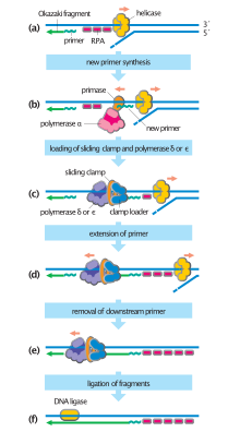
The RNA segments are first synthesized by primase and then elongated by DNA polymerase.[3] Then the DNA polymerase forms a protein complex with two primase subunits to form the alpha DNA Polymerase primase complex. Primase is one of the most error prone and slow polymerases.[3] Primases in organisms such as E. coli synthesize around 2000 to 3000 primers at the rate of one primer per second.[4] Primase also acts as a halting mechanism to prevent the leading strand from outpacing the lagging strand by halting the progression of the replication fork.[5] The rate determining step in primase is when the first phosphodiester bond is formed between two molecules of RNA.[3]
The replication mechanisms differ between different bacteria and viruses where the primase covalently link to helicase in viruses such as the T7 bacteriophage.[5] In viruses such as the herpes simplex virus (HSV-1), primase can form complexes with helicase.[6] The primase-helicase complex is used to unwind dsDNA (double-stranded) and synthesizes the lagging strand using RNA primers[6] The majority of primers synthesized by primase are two to three nucleotides long.[6]
Types
There are two main types of primase: DnaG found in most bacteria, and the AEP (Archaeo-Eukaryote Primase) superfamily found in archaean and eukaryotic primases. While bacterial primases (DnaG-type) are composed of a single protein unit (a monomer) and synthesize RNA primers, AEP primases are usually composed of two different primase units (a heterodimer) and synthesize two-part primers with both RNA and DNA components.[7] While functionally similar, the two primase superfamilies evolved independently of each other.
DnaG
The crystal structure of primase in E. coli with a core containing the DnaG protein was determined in the year 2000.[4] The DnaG and primase complex is cashew shaped and contains three subdomains.[4] The central subdomain forms a toprim fold which is made of a mixture five beta sheets and six alpha helices.[4][8] The toprim fold is used for binding regulators and metals. The primase uses a phosphotransfer domain for the transfer coordination of metals, which makes it distinct from other polymerases.[4] The side subunits contain a NH2 and COOH terminal made of alpha helixes and beta sheets.[4] The NH2 terminal interacts with a zinc binding domain and COOH-terminal region which interacts with DnaB-ID.[4]
The Toprim fold is also found in topoisomerase and mitochrondrial Twinkle primase/helicase.[8] Some DnaG-like (bacteria-like; InterPro: IPR020607) primases have been found in archaeal genomes.[9]
AEP
Eukaryote and archaeal primases tend to be more similar to each other, in terms of structure and mechanism, than they are to bacterial primases.[10][11] The archaea-eukaryotic primase (AEP) superfamily, which most eukaryal and archaeal primase catalytic subunits belong to, has recently been redefined as a primase-polymerase family in recognition of the many other roles played by enzymes in this family.[12] This classification also emphasizes the broad origins of AEP primases; the superfamily is now recognized as transitioning between RNA and DNA functions.[13]
Archaeal and eukaryote primases are heterodimeric proteins with one large regulatory (human PRIM2, p58) and one small catalytic subunit (human PRIM1, p48/p49).[2] The large subunit contains a N-terminal 4Fe–4S cluster, split out in some archaea as PriX/PriCT.[14] The large subunit is implicated in improving the activity and specificity of the small subunit. For example, removing the part corresponding to the large subunit in a fusion protein PolpTN2 results in a slower enzyme with reverse transcriptase activity.[13]
Multifunctional primases

The AEP family of primase-polymerases has diverse features beyond making only primers. In addition to priming DNA during replication, AEP enzymes may have additional functions in the DNA replication process, such as polymerization of DNA or RNA, terminal transfer, translesion synthesis (TLS), non-homologous end joining (NHEJ),[12] and possibly in restarting stalled replication forks.[15] Primases typically synthesize primers from ribonucleotides (NTPs); however, primases with polymerase capabilities also have an affinity for deoxyribonucleotides (dNTPs).[16][11] Primases with terminal transferase functionality are capable of adding nucleotides to the 3’ end of a DNA strand independently of a template. Other enzymes involved in DNA replication, such as helicases, may also exhibit primase activity.[17]
In eukaryotes and archaea
Human PrimPol (ccdc111[16]) serves both primase and polymerase functions, like many archaeal primases; exhibits terminal transferase activity in the presence of manganese; and plays a significant role in translesion synthesis[18] and in restarting stalled replication forks. PrimPol is actively recruited to damaged sites through its interaction with RPA, an adapter protein that facilitates DNA replication and repair.[15] PrimPol has a zinc finger domain similar to that of some viral primases, which is essential for translesion synthesis and primase activity and may regulate primer length.[18] Unlike most primases, PrimPol is uniquely capable of starting DNA chains with dNTPs.[16]
PriS, the archaeal primase small subunit, has a role in translesion synthesis (TLS) and can bypass common DNA lesions. Most archaea lack the specialized polymerases that perform TLS in eukaryotes and bacteria.[19] PriS alone preferentially synthesizes strings of DNA; but in combination with PriL, the large subunit, RNA polymerase activity is increased.[20]
In Sulfolobus solfataricus, the primase heterodimer PriSL can act as a primase, polymerase, and terminal transferase. PriSL is thought to initiate primer synthesis with NTPs and then switch to dNTPs. The enzyme can polymerize RNA or DNA chains, with DNA products reaching as long as 7000 nucleotides (7 kb). It is suggested that this dual functionality may be a common feature of archaeal primases.[11]
In bacteria
AEP multifutional primases also appear in bacteria and phages that infect them. They can display novel domain organizations with domains that bring even more functions beyond polymerization.[14]
Bacterial LigD (A0R3R7) is primarily involved in the NHEJ pathway. It has an AEP superfamily polymerase/primase domain, a 3'-phosphoesterase domain, and a ligase domain. It is also capable of primase, DNA and RNA polymerase, and terminal transferase activity. DNA polymerization activity can produce chains over 7000 nucleotides (7 kb) in length, while RNA polymerization produces chains up to 1 kb long.[21]
In viruses and plasmids
AEP enzymes are widespread, and can be found encoded in mobile genetic elements including virus/phages and plasmids. They either use them as a sole replication protein or in combination with other replication-associated proteins, such as helicases and, less frequently, DNA polymerases.[22] Whereas the presence of AEP in eukaryotic and archaeal viruses is expected in that they mirror their hosts,[22] bacterial viruses and plasmids also as frequently encode AEP-superfamily enzymes as they do DnaG-family primases.[14] A great diversity of AEP families has been uncovered in various bacterial plasmids by comparative genomics surveys.[14] Their evolutionary history is currently unknown, as these found in bacteria and bacteriophages appear too different from their archaeo-eukaryotic homologs for a recent horizontal gene transfer.[22]
MCM-like helicase in Bacillus cereus strain ATCC 14579 (BcMCM; Q81EV1) is an SF6 helicase fused with an AEP primase. The enzyme has both primase and polymerase functions in addition to helicase function. The gene coding for it is found in a prophage.[17] It bears homology to ORF904 of plasmid pRN1 from Sulfolobus islandicus, which has an AEP PrimPol domain.[23] Vaccinia virus D5 and HSV Primase are examples of AEP-helicase fusion as well.[12][6]
PolpTN2 is an Archaeal primase found in the TN2 plasmid. A fusion of domains homologous to PriS and PriL, it exhibits both primase and DNA polymerase activity, as well as terminal transferase function. Unlike most primases, PolpTN2 forms primers composed exclusively of dNTPs.[13] Unexpectedly, when the PriL-like domain was truncated, PolpTN2 could also synthesize DNA on the RNA template, i.e., acted as an RNA-dependent DNA polymerase (reverse transcriptase).[13]
Even DnaG primases can have extra functions, if given the right domains. The T7 phage gp4 is a DnaG primase-helicase fusion, and performs both functions in replication.[5]
References
- PMID 11301257.
- ^ PMID 25550159.
- ^ PMID 8655184.
- ^ PMID 10741967.
- ^ S2CID 3099842.
- ^ PMID 19028696.
- S2CID 17108681.
- ^ PMID 9722641.
- PMID 23324612.
- PMID 16027112.
- ^ PMID 15561142.
- ^ PMID 26109351.
- ^ PMID 24445805.
- ^ PMID 29198957.
- ^ PMID 24126761.
- ^ PMID 24207056.
- ^ PMID 21984415.
- ^ PMID 24682820.
- PMID 25646444.
- PMID 17158702.
- PMID 16095750.
- ^ PMID 27112572.
- S2CID 25123984.
