
Pulmonary artery
| Pulmonary artery | |
|---|---|
right ventricle | |
| Identifiers | |
| Latin | arteria pulmonalis |
| MeSH | D011651 |
| TA98 | A12.2.01.101 A12.2.01.201 |
| TA2 | 4077, 4091 |
| FMA | 66326 |
| Anatomical terminology] | |
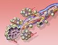
A pulmonary artery is an artery in the pulmonary circulation that carries deoxygenated blood from the right side of the heart to the lungs. The largest pulmonary artery is the main pulmonary artery or pulmonary trunk from the heart, and the smallest ones are the arterioles, which lead to the capillaries that surround the pulmonary alveoli.
Structure
The pulmonary arteries are blood vessels that carry systemic venous blood from the right ventricle of the heart to the microcirculation of the lungs. Unlike in other organs where arteries supply oxygenated blood, the blood carried by the pulmonary arteries is deoxygenated, as it is venous blood returning to the heart. The main pulmonary arteries emerge from the right side of the heart and then split into smaller arteries that progressively divide and become arterioles, eventually narrowing into the capillary microcirculation of the lungs where gas exchange occurs.[citation needed]
Pulmonary trunk
In order of blood flow, the pulmonary arteries start as the pulmonary trunk that leaves the fibrous pericardium (
The pulmonary trunk splits into the right and the left main pulmonary artery.[5] The left main pulmonary artery is shorter than the right,[1] passes behind and downwards the descending aorta and above the left main bronchus to the root of the left lung. Above, the left main pulmonary artery is connected to the concavity of the proximal descending aorta by the ligamentum arteriosum.[2] The right pulmonary artery pass across the midline of the body, below the carina of trachea, and comes in front of the right main bronchus.[1]
Branches

The left main pulmonary artery then divides into two lobar arteries, one for each lobe of the left lung.[6]
At the right root of the lung, it bifurcates into artery that supplies the right upper lobe of the lung, in front of the right upper lobe bronchus, and interlobar artery that supplies the right middle and inferior lobes of the lung, running together with bronchus intermedius.[1]
The right and left main pulmonary (lungs) arteries give off branches that supplies the corresponding
Development
The pulmonary arteries originate from the
By the third week of
During early development, the ductus arteriosus connects the pulmonary trunk and the aortic arch, allowing blood to bypass the lungs.[10]: 791
Function
The pulmonary artery carries deoxygenated blood from the
In contrast to the pulmonary arteries, the
Pressure
The pulmonary artery pressure (PA pressure) is a measure of the
Clinical significance
The pulmonary artery is relevant in a number of clinical states.
Pulmonary embolism refers to an embolus that lodges in the pulmonary circulation. This may arise from a
Several animal models have been utilized for investigating pulmonary artery related pathologies. Porcine model of pulmonary artery is the most frequently used and it was recently found that their mechanical properties vary with every subsequent branching.[20]
Additional images
-
 Image showing main pulmonary artery coursing ventrally to thetrachea, and the right pulmonary artery passes dorsally to the ascending aorta, while the left pulmonary artery passes ventrally to the descending aorta.
 -
 Pulmonary circuit
 -
 Transverse section of thorax, showing relations of pulmonary artery.
 -
 Pulmonary artery
 -
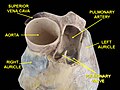 Pulmonary artery.Deep dissection.Anterior view.
 -
 CT scan of a normal lung, with different levels of pulmonary arteries.
 -
 Bronchial anatomy


.jpg)


See also
- Pulmonary artery sling
- Rasmussen's aneurysm
- Pulmonary vein
- Pulmonary circulation
- Bronchial artery
- Bronchial vein
- Brachial artery
- Brachial vein
- Coronary artery
- Coronary sinus
- Coronary vein)
- Pulmonary valve
References
- ^ ISBN 9780702029714.
- ^ ISBN 9789350252758.
- PMID 10211060.
- PMID 22178898.
- ^ "Pulmonary Vasculature". University of Virginia School of Medicine. 2013. Retrieved 2017-06-24.
- PMID 30057869.
- ^ a b c "Pulmonary Artery Anatomy". University of Virginia School of Medicine. 2013. Retrieved 2017-06-24.
- PMID 18686729.
- ^ ISBN 9780443068119.
- ^ a b Braunwald E (1992). Heart Disease: A Textbook of Cardiovascular Medicine (Fourth ed.). Philadelphia: W.B. Sanders.
- ^ "22.4 Gas Exchange – Anatomy and Physiology". opentextbc.ca. Archived from the original on 2020-10-19. Retrieved 2019-05-22.
- ^ "Exchanging Oxygen and Carbon Dioxide – Lung and Airway Disorders". MSD Manual Consumer Version. Retrieved 2019-05-22.
- ^ ISBN 978-0-7020-3084-0.
- ^ "Normal Hemodynamic Parameters – Adult" (PDF). Edwards Lifesciences LLC. Archived from the original (PDF) on 2010-11-10.
- S2CID 26693444.
- PMID 30382495.
- PMID 5865788. Retrieved 15 January 2022.
- PMID 32007361.
- ^ Jones J, et al. "Saddle pulmonary embolism". Radiopaedia. Retrieved 2017-10-08.
- PMID 33814554.
External links
- Anatomy photo:20:01-0106 at the SUNY Downstate Medical Center – "Heart: The Pericardial sac and Great vessels"
- Anatomy photo:20:07-0105 at the SUNY Downstate Medical Center – "Heart: Openings of Great Vessels into the Pericardial Sac"
- Anatomy figure: 19:05-06 at Human Anatomy Online, SUNY Downstate Medical Center – "right lung"
- Anatomy figure: 19:06-02 at Human Anatomy Online, SUNY Downstate Medical Center – "Mediastinal surface of the left lung"
- Histology image: 13802loa – Histology Learning System at Boston University
| National | |
|---|---|
| Other | |